. 2008 Jan 10;57(1):108-20.
doi: 10.1016/j.neuron.2007.11.024.

Long-term potentiation selectively expressed by NMDA receptors at hippocampal mossy fiber synapses

Affiliations

Long-term potentiation selectively expressed by NMDA receptors at hippocampal mossy fiber synapses

Hyung-Bae Kwon et al. Neuron. .

Abstract

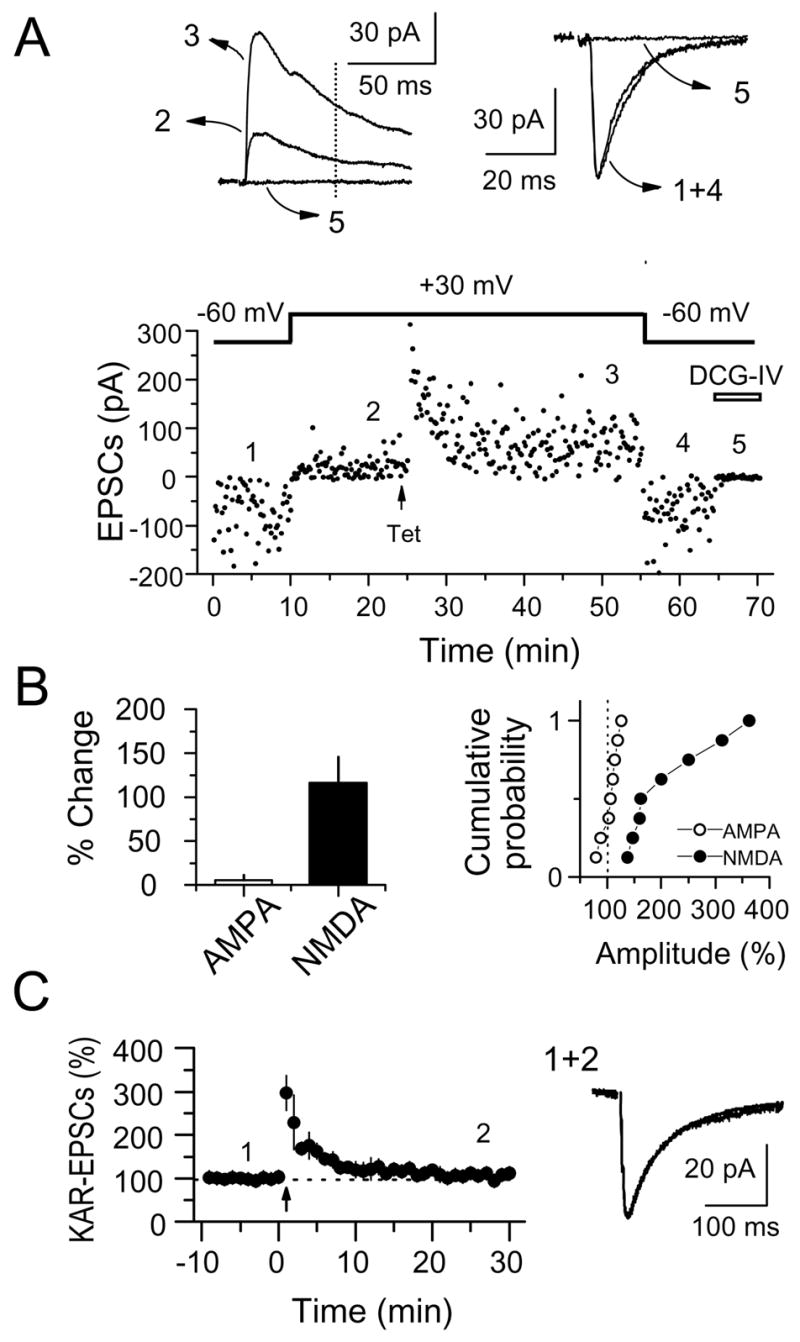
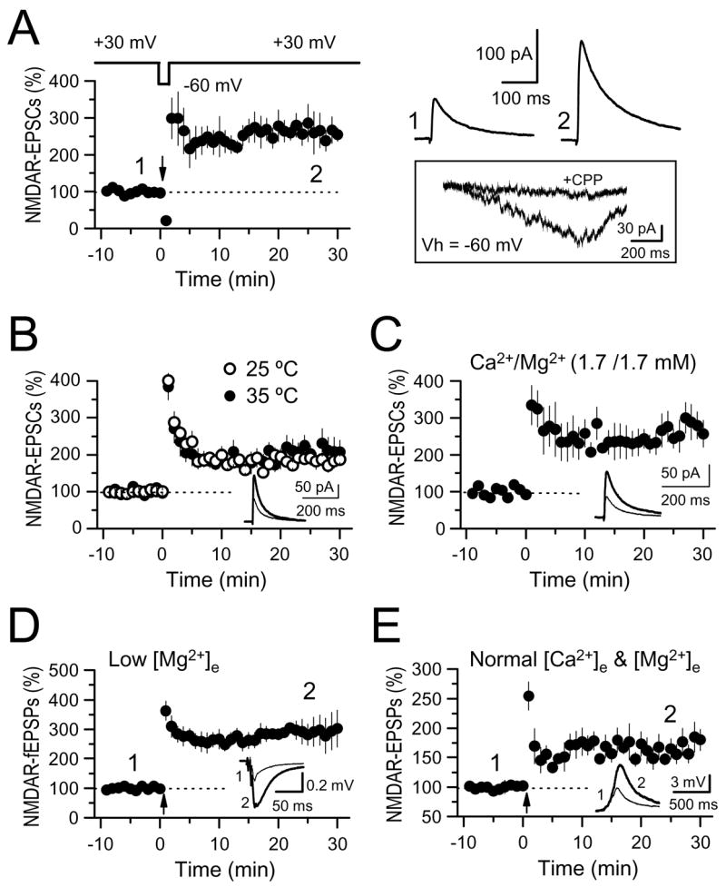
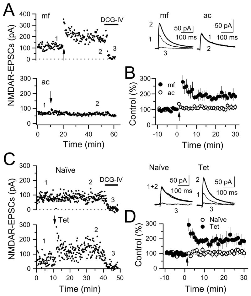
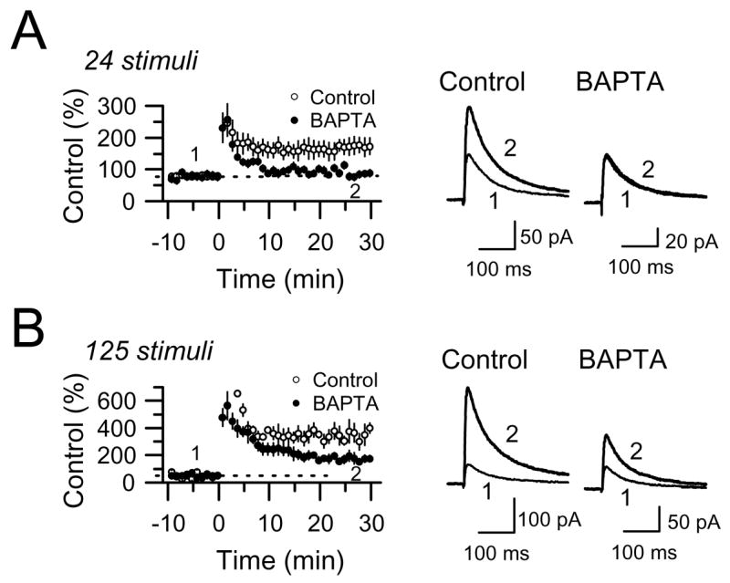
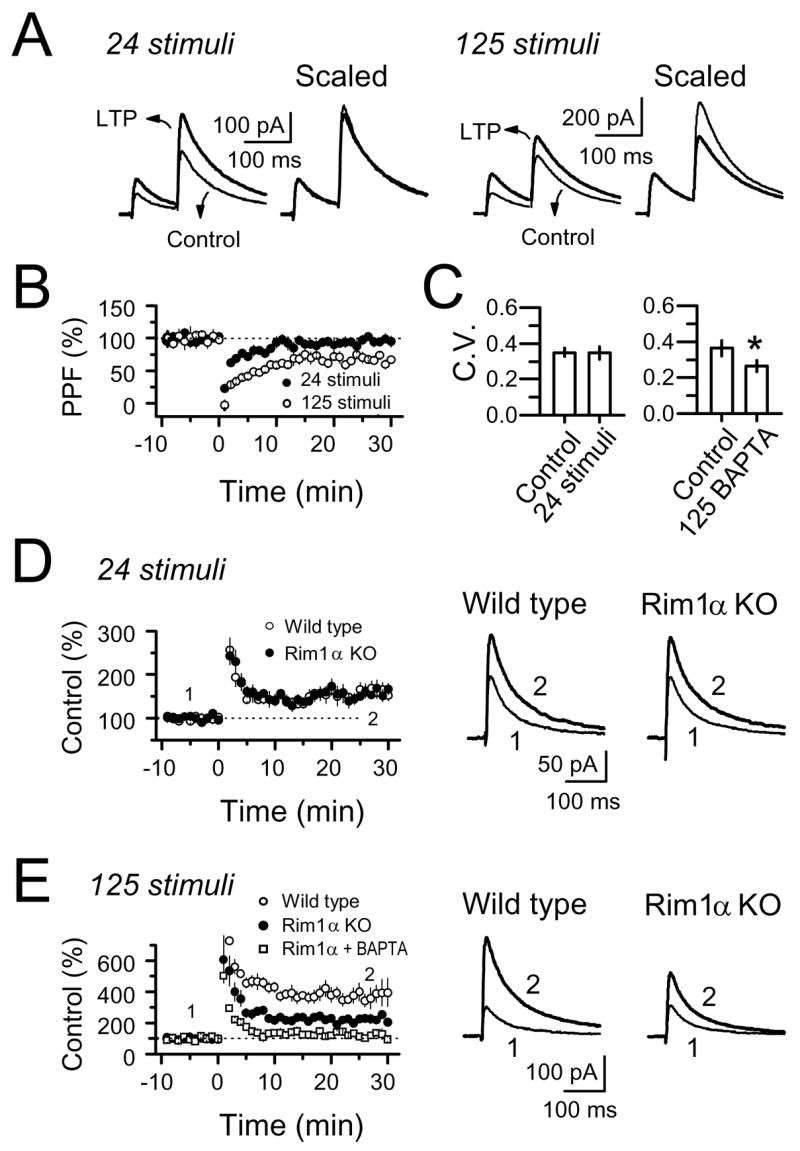
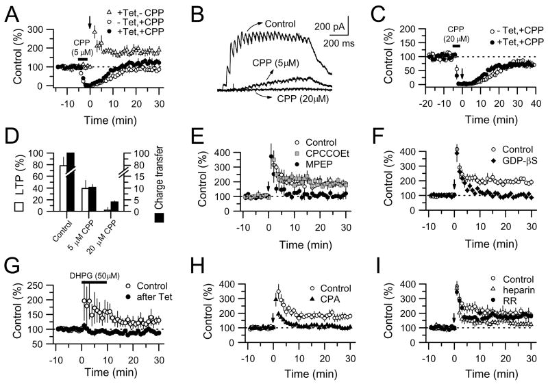
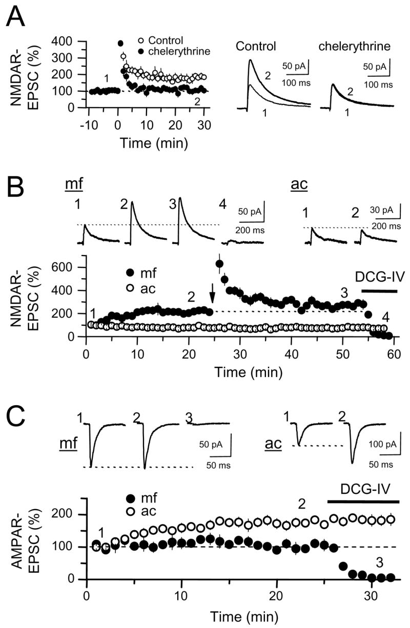
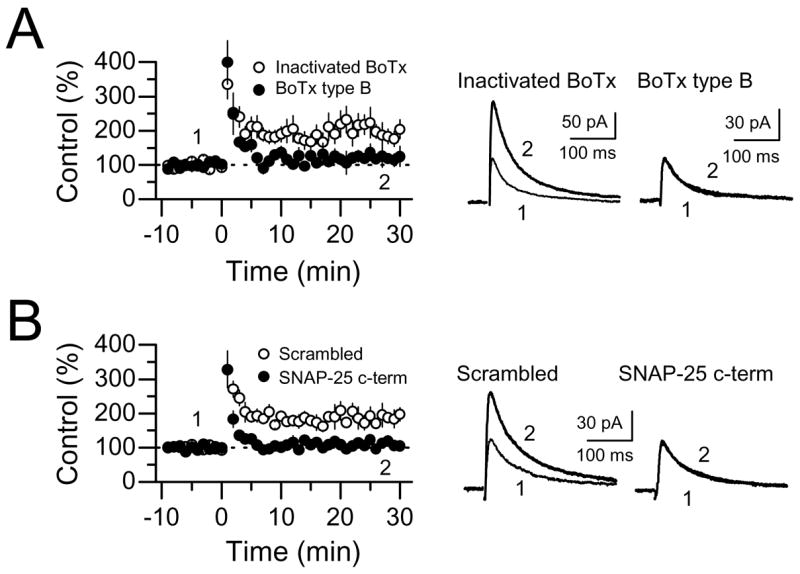
The mossy fiber to CA3 pyramidal cell synapse (mf-CA3) provides a major source of excitation to the hippocampus. Thus far, these glutamatergic synapses are well recognized for showing a presynaptic, NMDA receptor-independent form of LTP that is expressed as a long-lasting increase of transmitter release. Here, we show that in addition to this "classical" LTP, mf-CA3 synapses can undergo a form of LTP characterized by a selective enhancement of NMDA receptor-mediated transmission. This potentiation requires coactivation of NMDA and mGlu5 receptors and a postsynaptic calcium rise. Unlike classical LTP, expression of this mossy fiber LTP is due to a PKC-dependent recruitment of NMDA receptors specifically to the mf-CA3 synapse via a SNARE-dependent process. Having two mechanistically different forms of LTP may allow mf-CA3 synapses to respond with more flexibility to the changing demands of the hippocampal network.


Figures

Figure 1
Figure 1
Selective LTP of NMDAR-EPSCs at mf-CA3 synapses (NMDAR-mfLTP). (A) Representative experiment in which both NMDAR- and AMPAR-EPSCs from a CA3 pyramidal cell were monitored over time. LTP was induced by a single tetanus (Tet) of 24 stimuli at 25 Hz. At the end of the experiment, the mGluR2 agonist DCG-IV (1 μM) was applied to confirm that EPSCs were indeed mediated by mossy fibers. NMDAR-EPSC amplitudes were measured 50 ms post-stimulus (vertical dotted line) to avoid the contribution of the AMPAR-mediated component. Averaged sample traces taken at times indicated by numbers are shown on top. (B) Summary plots showing the selective potentiation of NMDAR-EPSCs but not AMPAR-EPSCs (8 cells). (C) Same induction protocol (24 pulses at 25 Hz) failed to increase KAR-EPSCs (4 cells). Average traces before and after tetanus are shown on the right.
Figure 2
Figure 2
NMDAR-mfLTP can be induced under more physiological conditions. (A) mf NMDAR-EPSCs were monitored at Vh = +30 mV, but the induction tetanus was delivered at Vh = β60 mV (4 cells). Averaged sample traces taken at times indicated by numbers are shown on the top right. Below that (box) are representative traces of the NMDAR-mediated inward current induced by the tetanus (24 stimuli, 25 Hz) before and after application of 20 μM CPP. Stimulation artifacts were deleted for clarity. (B) Summary plots showing that the magnitude of NMDAR-mf LTP obtained at 25°C (6 cells) and 35°C (6 cells) is virtually identical. (C) Summary plots showing NMDAR-mfLTP while using 1.7 mM Ca2+ and 1.7 mM Mg2+ in the extracellular solution (5 cells). (D) NMDAR-mfLTP was induced while monitoring field potential amplitude (NMDAR-fEPSPs) in a 4.0 mM Ca2+, 0.1 mM Mg2+ extracellular solution (3 slices). Each fEPSP was induced with a burst of 3 stimuli at 200 Hz. (E) NMDAR-mfLTP was induced while recording NMDAR-EPSPs in current clamp mode, and in the presence of more physiological extracellular concentrations of Ca2+ and Mg2+ (2.5 and 1.3 mM, respectively) (4 cells). Each EPSP was induced with a burst of 5 stimuli at 25 Hz. Insets of B, C, D and E: averaged sample traces from before and after NMDAR-mfLTP are superimposed.
Figure 3
Figure 3
NMDAR-mfLTP is input-specific. (A) Time-course of a representative experiment in which NMDAR-EPSCs evoked by alternating stimulation of mfs (mf, top panel) and associational-commissural fibers (ac, bottom panel) were monitored in the same CA3 pyramidal cell. Tetanic stimulation (vertical arrow: 24 stimuli, 25 Hz) induced LTP at mf-CA3 synapses only. Averaged sample traces taken at times indicated by numbers (upper right). (B) Summary plot of 5 cells recorded as in A showing robust LTP of NMDAR-EPSCs at mf-CA3 synapses only. (C) Representative experiment in which NMDAR-EPSCs were evoked by alternatively stimulating two independent mf pathways. At the time point indicated by the vertical arrow, one pathway received a tetanus of 24 stimuli at 25 Hz (Tet) whereas the naïve pathway served as control. Averaged sample traces taken at times indicated by numbers (right). (D) Summary plot of 4 cells as recorded in C showing that NMDAR-mfLTP was induced at mf-CA3 synapses that received the induction tetanus but not at naïve synapses.
Figure 4
Figure 4
Role of postsynaptic Ca2+ in the induction of NMDAR-mfLTP. (A) Loading CA3 pyramidal cells with 20 mM BAPTA abolished NMDAR-mfLTP induced by 24 stimuli at 25 Hz (6 cells in BAPTA and 6 control cells). (B) A longer induction tetanus (125 stimuli, 25 Hz) triggered a larger potentiation with BAPTA-sensitive and BAPTA-insensitive components (6 cells in BAPTA and 6 control cells).
Figure 5
Figure 5
NMDAR-mfLTP (24 stimuli, 25 Hz) differs from the classical presynaptic form of LTP (125 stimuli, 25 Hz). LTP induced by 24 stimuli had no lasting effect on paired-pulse facilitation (PPF) or coefficient of variation (CV), but LTP induced by 125 stimuli significantly reduced both. (A) Averaged traces before and after LTP induced by either 24 or 125 stimuli. (B) PPF. (C) CV. (D) RIM1α deletion had no effect on NMDAR-mfLTP (4 WT mice and 4 RIM1α KO mice). (E) A longer tetanus (125 stimuli) induced robust mfLTP which included a RIM1α-dependent component (presynaptic mfLTP) and a RIM1α-independent component (NMDAR-mfLTP) (4 WT mice and 4 RIM1α KO mice). This latter component was abolished by BAPTA (4 cells/2 mice).
Figure 6
Figure 6
NMDAR-mfLTP requires the coactivation of NMDA and mGluR5 receptors, and Ca2+ release from postsynaptic IP3-sensitive stores. (A) Transiently blocking NMDARs during tetanus (7 cells) inhibited induction of NMDAR-mfLTP. The NMDAR antagonist CPP (5 μM, horizontal bar) was bath applied for 4 min to slices receiving the tetanus (Tet) and also to naïve slices (4 cells) to map the rate of CPP washout. NMDAR-mfLTP in control slices (7 cells) is superimposed for comparison. (B) Representative NMDAR-mediated currents induced by the tetanus under control conditions and in the presence of 5 μM and 20 μM CPP. Note that 5 μM CPP, a competitive antagonist, was insufficient to completely block these currents, whereas 20 μM CPP produced a complete block. (C) Same procedure as in panel (A), but using enough CPP (20 μM) to completely block NMDAR current during the tetanus (5 cells). (D) Summary graph showing that while incomplete blockade of NMDARs (measured as charge transfer) during tetanus application reduced NMDAR-mfLTP only partially, full blockade of NMDARs abolished NMDAR-mfLTP completely. White bars indicate the magnitude of LTP, black bars indicate charge transfer normalized to control conditions (e.g. in the absence of CPP). (E) Activation of mGluR5, but not mGluR1, is required for NMDAR-mfLTP. Summary plot comparing three experimental groups in which the induction tetanus was delivered in the presence of the mGluR5 antagonist MPEP (4 μM, 6 cells), the mGluR1 antagonist CPCCOEt (100 μM, 4 cells), or in interleaved control slices (n = 6 cells). (F) Effects of GDP-βS (2 mM) postsynaptic loading on NMDAR-mfLTP. GDP-βS was allowed to diffuse into CA3 cells (n = 4) at least for 30 minutes before tetanus. For comparison, NMDAR-mfLTP elicited in interleaved control experiments (n = 4) is superimposed. (G) Summary plots showing that bath application of 50 μM DHPG (in the presence of 100 μM CPCCOEt) induced weak LTP of mf NMDAR-EPSCs (Control, 8 cells). In separate set of experiments, this potentiation was occluded by prior induction of NMDAR-mfLTP (after Tet, 6 cells). (H) NMDAR-mfLTP requires Ca2+ release from IP3-sensitive Ca2+ stores. Summary plots comparing the magnitude of NMDAR-mfLTP in hippocampal slices treated with cyclopiazonic acid (CPA, 5 cells) and in interleaved control slices (5 cells). Test hippocampal slices were incubated in 30 μM CPA for at least 30 min before and continuously during recordings. (I) Summary plots showing the effects of the IP3-receptor blocker heparin (2.5 mg/ml) or the ryanodine receptor blocker ruthenium red (RR) (20 μM) on NMDAR-mfLTP. Heparin (5 cells) and RR (4 cells) were added to the recording pipette.
Figure 7
Figure 7
PKC activation is required to induce NMDAR-mfLTP, and it is sufficient to potentiate NMDAR-mfEPSCs. (A) Loading CA3 pyramidal cells (n = 6) with the PKC blocker chelerythrine (10 μM) abolished NMDAR-mfLTP. (B) Loading CA3 pyramidal cells with PKM, a constitutively active fragment of PKC, potentiated NMDAR-mfEPSCs (4 cells). Note that PKM potentiated mossy fiber (mf) but not associational-commissural (ac) NMDAR-EPSCs. Once PKM-mediated potentiation stabilized, subsequent tetanus (24 stimuli, 25 Hz) produced only a weak NMDAR-mfLTP, likely due to occlusion. Averaged sample traces taken at the times indicated by numbers are inset above. (C) Same procedure as in (B) while monitoring AMPAR-EPSCs. PKM potentiated the AMPAR-mediated transmission at ac-CA3 but not mf-CA3 synapses (7 cells).
Figure 8
Figure 8
NMDAR-mfLTP requires SNARE-dependent exocytosis. (A) Summary plots showing the effects of the recombinant light chain of botulinum neurotoxin (BoTx) type B on NMDAR-mfLTP (5 cells). Heat-inactivated BoTx (100 ng/ml) was used as control (6 cells). Both BoTx and heat-inactivated BoTx were loaded into the CA3 pyramidal cell, and allowed to perfuse for at least 30 min before tetanic stimulation. (B) Summary plots comparing the effects of a short SNAP-25 interfering peptide (SNAP-25 c-term)(5 cells) and a scrambled peptide on NMDAR-mfLTP (6 cells). Averaged sample traces are shown on the right side.

Comment in

Similar articles

Cited by

References

    1. Abraham WC, Bear MF. Metaplasticity: the plasticity of synaptic plasticity. Trends Neurosci. 1996;19:126–130. - PubMed
    1. Aniksztejn L, Ben-Ari Y. Expression of LTP by AMPA and/or NMDA receptors is determined by the extent of NMDA receptors activation during the tetanus. J Neurophysiol. 1995;74:2349–2357. - PubMed
    1. Barria A, Malinow R. Subunit-specific NMDA receptor trafficking to synapses. Neuron. 2002;35:345–353. - PubMed
    1. Bashir ZI, Alford S, Davies SN, Randall AD, Collingridge GL. Long-term potentiation of NMDA receptor-mediated synaptic transmission in the hippocampus. Nature. 1991;349:156–158. - PubMed
    1. Bayazitov I, Kleschevnikov A. Afferent high strength tetanizations favour potentiation of the NMDA vs. AMPA receptor-mediated component of field EPSP in CA1 hippocampal slices of rats. Brain Res. 2000;866:188–196. - PubMed

Publication types

MeSH terms

Substances