

Structure of the human protein kinase MPSK1 reveals an atypical activation loop architecture
- PMID: 18184589
- PMCID: PMC2194165
- DOI: 10.1016/j.str.2007.10.026
Structure of the human protein kinase MPSK1 reveals an atypical activation loop architecture
Abstract
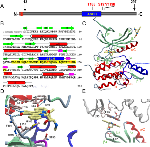
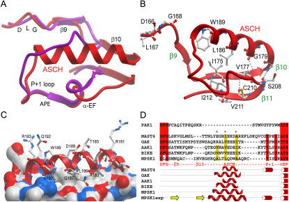
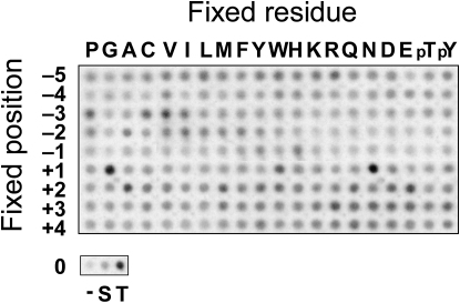
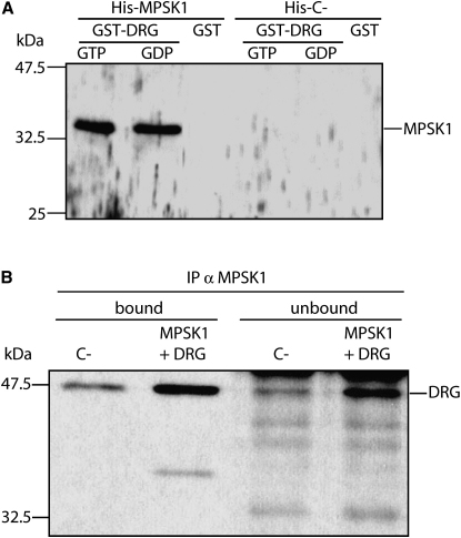
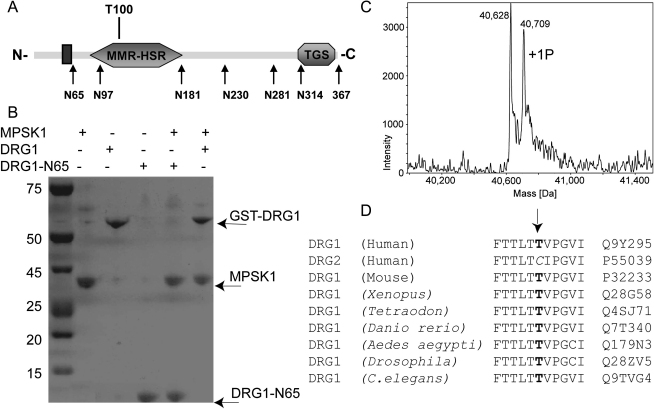
The activation segment of protein kinases is structurally highly conserved and central to regulation of kinase activation. Here we report an atypical activation segment architecture in human MPSK1 comprising a beta sheet and a large alpha-helical insertion. Sequence comparisons suggested that similar activation segments exist in all members of the MPSK1 family and in MAST kinases. The consequence of this nonclassical activation segment on substrate recognition was studied using peptide library screens that revealed a preferred substrate sequence of X-X-P/V/I-phi-H/Y-T*-N/G-X-X-X (phi is an aliphatic residue). In addition, we identified the GTPase DRG1 as an MPSK1 interaction partner and specific substrate. The interaction domain in DRG1 was mapped to the N terminus, leading to recruitment and phosphorylation at Thr100 within the GTPase domain. The presented data reveal an atypical kinase structural motif and suggest a role of MPSK1 regulating DRG1, a GTPase involved in regulation of cellular growth.
Figures
Similar articles
-
Serine/Threonine Protein Kinase STK16.Int J Mol Sci. 2019 Apr 10;20(7):1760. doi: 10.3390/ijms20071760. Int J Mol Sci. 2019. PMID: 30974739 Free PMC article. Review.
-
Crystal structure of the ALK (anaplastic lymphoma kinase) catalytic domain.Biochem J. 2010 Sep 15;430(3):425-37. doi: 10.1042/BJ20100609. Biochem J. 2010. PMID: 20632993
-
Catalytically active MAP KAP kinase 2 structures in complex with staurosporine and ADP reveal differences with the autoinhibited enzyme.Structure. 2003 Jun;11(6):627-36. doi: 10.1016/s0969-2126(03)00092-3. Structure. 2003. PMID: 12791252
-
Characterization of a domain that transiently converts class 2 DYRKs into intramolecular tyrosine kinases.Sci Signal. 2010 Mar 2;3(111):ra16. doi: 10.1126/scisignal.2000579. Sci Signal. 2010. PMID: 20197545
-
A conserved Gbeta binding (GBB) sequence motif in Ste20p/PAK family protein kinases.Biol Chem. 2000 May-Jun;381(5-6):427-31. doi: 10.1515/BC.2000.055. Biol Chem. 2000. PMID: 10937873 Review.
Cited by
-
A kinase cascade on the yeast lysosomal vacuole regulates its membrane dynamics: conserved kinase Env7 is phosphorylated by casein kinase Yck3.J Biol Chem. 2020 Aug 21;295(34):12262-12278. doi: 10.1074/jbc.RA119.012346. Epub 2020 Jul 9. J Biol Chem. 2020. PMID: 32647006 Free PMC article.
-
Structural coupling of SH2-kinase domains links Fes and Abl substrate recognition and kinase activation.Cell. 2008 Sep 5;134(5):793-803. doi: 10.1016/j.cell.2008.07.047. Cell. 2008. PMID: 18775312 Free PMC article.
-
Insights into protein kinase regulation and inhibition by large scale structural comparison.Biochim Biophys Acta. 2010 Mar;1804(3):429-32. doi: 10.1016/j.bbapap.2009.10.013. Epub 2009 Oct 22. Biochim Biophys Acta. 2010. PMID: 19854302 Free PMC article. Review.
-
A Serine/Threonine Kinase 16-Based Phospho-Proteomics Screen Identifies WD Repeat Protein-1 As A Regulator Of Constitutive Secretion.Sci Rep. 2018 Aug 29;8(1):13049. doi: 10.1038/s41598-018-31426-1. Sci Rep. 2018. PMID: 30158666 Free PMC article.
-
Developmentally regulated GTPases: structure, function and roles in disease.Cell Mol Life Sci. 2021 Dec;78(23):7219-7235. doi: 10.1007/s00018-021-03961-0. Epub 2021 Oct 19. Cell Mol Life Sci. 2021. PMID: 34664086 Free PMC article. Review.
References
-
- Aimes R.T., Hemmer W., Taylor S.S. Serine-53 at the tip of the glycine-rich loop of cAMP-dependent protein kinase: role in catalysis, P-site specificity, and interaction with inhibitors. Biochemistry. 2000;39:8325–8332. - PubMed
-
- Berson A.E., Young C., Morrison S.L., Fujii G.H., Sheung J., Wu B., Bolen J.B., Burkhardt A.L. Identification and characterization of a myristylated and palmitylated serine/threonine protein kinase. Biochem. Biophys. Res. Commun. 1999;259:533–538. - PubMed
-
- Bertrand J.A., Thieffine S., Vulpetti A., Cristiani C., Valsasina B., Knapp S., Kalisz H.M., Flocco M. Structural characterization of the GSK-3β active site using selective and non-selective ATP-mimetic inhibitors. J. Mol. Biol. 2003;333:393–407. - PubMed
-
- Bossemeyer D. The glycine-rich sequence of protein kinases: a multifunctional element. Trends Biochem. Sci. 1994;19:201–205. - PubMed
-
- Brown N.R., Noble M.E., Lawrie A.M., Morris M.C., Tunnah P., Divita G., Johnson L.N., Endicott J.A. Effects of phosphorylation of threonine 160 on cyclin-dependent kinase 2 structure and activity. J. Biol. Chem. 1999;274:8746–8756. - PubMed
Publication types
MeSH terms
Substances
Grants and funding
LinkOut - more resources
Full Text Sources
Molecular Biology Databases
