Review
. 2008 Jun 6;5(23):575-93.
doi: 10.1098/rsif.2007.1281.

Hydrodynamic aspects of fish olfaction

Affiliations
Review

Hydrodynamic aspects of fish olfaction

Jonathan P L Cox. J R Soc Interface. .

Abstract

Flow into and around the olfactory chamber of a fish determines how odorant from the fish's immediate environment is transported to the sensory surface (olfactory epithelium) lining the chamber. Diffusion times in water are long, even over comparatively short distances (millimetres). Therefore, transport from the external environment to the olfactory epithelium must be controlled by processes that rely on convection (i.e. the bulk flow of fluid). These include the beating of cilia lining the olfactory chamber and the relatively inexpensive pumping action of accessory sacs. Flow through the chamber may also be induced by an external flow. Flow over the olfactory epithelium appears to be laminar. Odorant transfer to the olfactory epithelium may be facilitated in several ways: if the olfactory organs are mounted on stalks that penetrate the boundary layer; by the steep velocity gradients generated by beating cilia; by devices that deflect flow into the olfactory chamber; by parallel arrays of olfactory lamellae; by mechanical agitation of the chamber (or olfactory stalks); and by vortices. Overall, however, our knowledge of the hydrodynamics of fish olfaction is far from complete. Several areas of future research are outlined.


Figures

Figure 1
Figure 1
Photograph of the head of a preserved northern pike (Esox lucius) specimen (total length 17 cm), lateral (l.h.s.) aspect. Boxed region highlights the anterior (A) and posterior (P) nostrils. Scale bar, 1 cm. Inset: boxed region from the main photograph. Scale bar, 1 mm. Photograph copyright © Natural History Museum, London (specimen catalogue number BMNH 1963.4.26: 1–2).
Figure 2
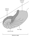
Figure 2
Schematic showing the main features of the olfactory organ of a northern pike (longitudinal cross section). The olfactory chamber is shaded light grey. This particular chamber includes an internal flap (F) believed to direct flow onto the sensory area (Burne 1909, p. 629). The latter is located between the low ridges and at the centre of the radial arrangement formed by the ridges. In fact, the ridge system is more complicated than that shown, with minor ridges lying between major ridges and transverse ridges connecting the major and minor ridges, giving a cobweb-like structure, with the sensory region occupying the spaces in the cobweb (Holl 1965, pp. 738–740). Schematic based on text-fig. 198 of Burne (1909) and fig. 21 of Teichmann (1954).
Figure 3
Figure 3
Photograph of the preserved head of a garpike (Belone belone) highlighting the triangular olfactory pit (boxed region). The olfactory epithelium coats both boss and pit (Theisen et al. 1980, fig. 2d). Scale bar, 1 cm. Inset: boxed region from the main photograph. Scale bar, 250 μm. Photographs copyright © Natural History Museum, London (specimen catalogue number BMNH 2005.4.27: 24–30).
Figure 4
Figure 4
Photograph of a live blackspotted puffer (A. nigropunctatus, normal phase). The olfactory organs are the pair of dark, forked structures lying within the boxed region. Inset: boxed region from the main photograph. Scale bar, approximately 3 mm. Photograph courtesy of Bristol Zoo Gardens, UK.
Figure 5
Figure 5
Three different types of olfactory lamellar array. (a) Longitudinal array of olfactory lamellae. (i) Photograph of lateral aspect of head of a preserved specimen of a male anglerfish (Linophryne species, total length 22 mm). Scale bar, 1 mm. Photograph copyright © Natural History Museum, London (specimen catalogue number BMNH 2004.11.6.44). (ii) Plan view of olfactory chamber. (b) Rosette. (i) Electron micrograph of olfactory chamber of goldfish (Carassius auratus). Scale bar, 0.3 mm. Reprinted with permission from Wiley-Liss, Inc., a subsidiary of John Wiley & Sons, Inc. from Hansen et al. (2004). Copyright © John Wiley & Sons, Inc. 2004. (ii) Plan view. (c) Elongated rosette. (i) Electron micrograph of the olfactory chamber of a European eel (Anguilla anguilla). Note that the raphe is gently curved rather than straight. Scale bar, 1 mm. Reproduced with permission from Springer Science+Business Media, LLC from Hansen & Zielinski (2005). Copyright © Springer Science+Business Media, LLC 2006. (ii) Plan view of olfactory chamber. A and P, outlines/positions of the anterior and posterior nostrils, respectively; d, distance between successive olfactory lamellae (referred to here as the depth of the olfactory lumen). The arrows in (b,c) highlight the rounded fin-like extremities of the lamellae, potential candidates for shedding tip vortices (§8).
Figure 6
Figure 6
Comparison of velocity profiles for fully developed flow between two wide but closely spaced parallel plates (P) and within a circular pipe (C). Flow between the parallel plates is, on the average, closer to the walls of the channel (CH) than in a circular pipe, given that the channels are of the same length, the diameter of the circular pipe is equal to the perpendicular distance between the two parallel plates and the fluid flowing through them has the same viscosity. The vertical lines below dP and dC show the average distances of the flow from the wall of the parallel plate channel and the circular pipe, respectively. Arrow, direction of flow; filled circles, the velocity at the wall is zero (the no-slip condition; Vogel 1994, pp. 18–20).
Figure 7
Figure 7
Boundary layers associated with swimming fish. The boundary layers indicated (shaded) are highly schematic, and are of course shown in two dimensions only. The relative motion of the fish with respect to the water (arrow) in each case is from right to left. (a) Profile of the head of a haddock (Melannogrammus aeglefinus) from a drawing (Wheeler 1969, p. 277). Approximate location of olfactory organ indicated by the filled oval. (b) Profile of the head of a bichir (Polypterus endlicheri) based on video footage of a swimming fish at Bristol Zoo Gardens, UK. Only the left-hand tubular anterior nostril is apparent (black). (c) Profile of the head of a blackspotted puffer taken from video footage of a swimming fish at Bristol Zoo Gardens, UK. Only the left-hand olfactory organ is apparent (black). Diagrams not to scale.
Figure 8
Figure 8
Schematic longitudinal cross section through the olfactory organ of an ‘oviparous cyprinodont’ fish (Zeiske 1974), a group to which the striped panchax belongs. Based on fig. 3a of Kux et al. (1988), the same cross section through the green swordtail is likely to be similar. Adapted from fig. 1 of Zeiske (1974).
Figure 9
Figure 9
Main pictures: photographs of the head of a preserved specimen of a sterlet (Acipenser ruthenus, total length approx. 35 cm). (a) Lateral view, showing the anterior and posterior nostrils of left-hand olfactory organ. Inset: photograph of the anterior and posterior nostrils of the olfactory organ of a lake sturgeon (Acipenser fulvescens, total length approx. 25 cm), highlighting the well-rounded rim of the anterior nostril (arrowhead). The posterior nostril of this particular organ is, unusually for sturgeons, partially occluded by a flap of skin (F); the posterior nostril of the other olfactory organ of this specimen was the more typical oval hole. Scale bar, 5 mm. (b) Dorsal view. Note the protruding posterior nostril. Scale bar, 1 cm. Scale for both (a,b) is the same. A, anterior nostril; P, posterior nostril. Photographs courtesy of the Natural History Museum (specimen catalogue numbers BMNH 1896.10.3.53-54 (A. ruthenus) and BMNH 1963.10.28.5 (A. fulvescens)).
Figure 10
Figure 10
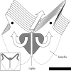
Schematic of part of the elongated olfactory rosette of the channel catfish (Ictalurus punctatus). Lamellae are attached to the raphe and the floor and sides of the olfactory chamber (dashed lines). The roof of the chamber is not shown in the main picture but is shown in the inset. The free edges of the lamellae protrude into the channel (grey in the inset) above the array. There is no information in the literature on the nature of this channel, though based on the anatomy of other fishes with similar olfactory organs (e.g. the European eel (Teichmann 1959, p. 241) and the catfish, Wallago attu (Ojha & Kapoor 1972, pp. 108–110)), the channel is probably narrow. The arrow above the array indicates the direction of flow in this channel. The sensory areas of the two nearest lamellar faces are shown in grey (only the sensory area on the nearest pair of lamellar faces is shown, for convenience); the remaining lamellar area is predominantly occupied by kinociliated cells. The curved arrows indicate the approximate direction of the flow over the lamellae. Again, there is no information in the literature on the direction of the flow over the olfactory lamellae of the channel catfish, and the direction shown here is an assumption based on the location of the kinociliated cells. Flow is only shown on the nearest pair of lamellar faces, but will be similarly directed over the other lamellar faces in the array. The fin-like dorsal regions (filled circles) of each lamella are potential candidates for shedding tip vortices (§8). Scale bar, 0.5 mm. The schematic is based on fig. 1 of Erickson & Caprio (1984).
Figure 11
Figure 11
Schematic showing how water expelled from a single accessory sac (large arrow) could cause a vortex (circular arrow) at the back of the olfactory chamber of a fish, leading to enhanced odorant transfer and possible entrainment of fluid through the anterior nostril (long arrow). Adapted from fig. 1 of Zeiske (1974); see also figure 8.

Similar articles

Cited by

References

    1. Alexander R.McN. Structure and function in the catfish. J. Zool. 1965;148:88–152.
    1. Alioto T.S, Ngai J. The odorant receptor repertoire of teleost fish. BMC Genomics. 2005;6:173. doi: 10.1186/1471-2164-6-173. - DOI - PMC - PubMed
    1. Anderson E.J, McGillis W.R, Grosenbaugh M.A. The boundary layer of swimming fish. J. Exp. Biol. 2001;204:81–102. - PubMed
    1. Anon. Fish images provide net benefit to researchers. Nature. 2006;440:396–397. doi: 10.1038/440396a. - DOI
    1. Atema J. Distribution of chemical stimuli. In: Atema J, Fay R.R, Popper A.N, Tavolga W.N, editors. Sensory biology of aquatic animals. Springer; New York, NY: 1988. pp. 29–56.

Publication types

LinkOut - more resources