. 2008 Mar;82(6):2989-99.
doi: 10.1128/JVI.01906-07. Epub 2008 Jan 9.

Envelope protein palmitoylations are crucial for murine coronavirus assembly

Affiliations

Envelope protein palmitoylations are crucial for murine coronavirus assembly

Joseph A Boscarino et al. J Virol. 2008 Mar.

Abstract

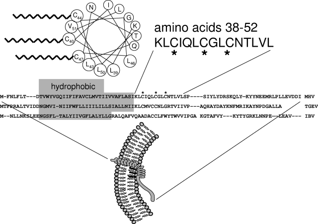
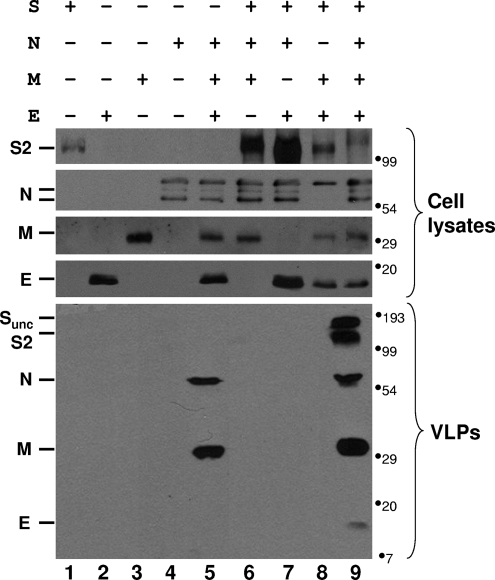
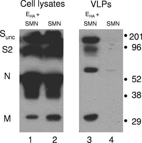
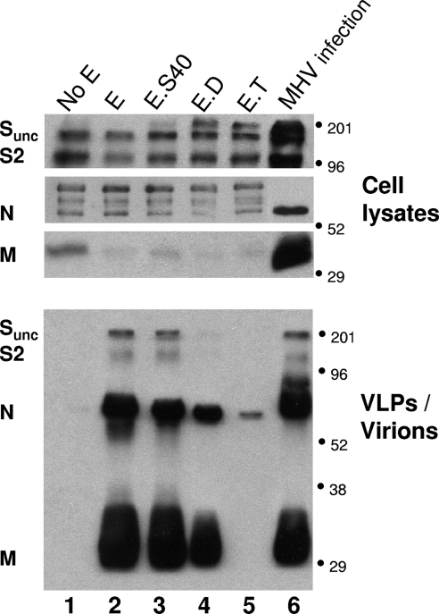
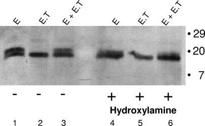
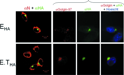
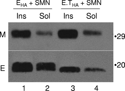
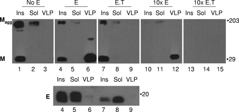
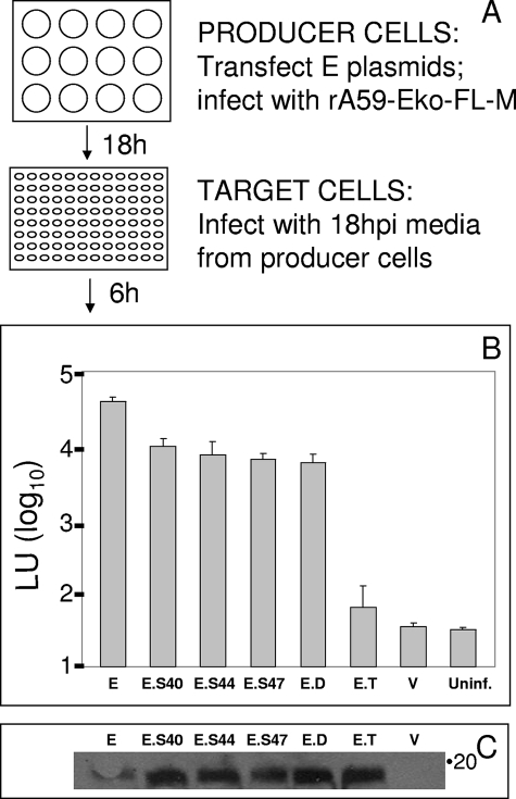
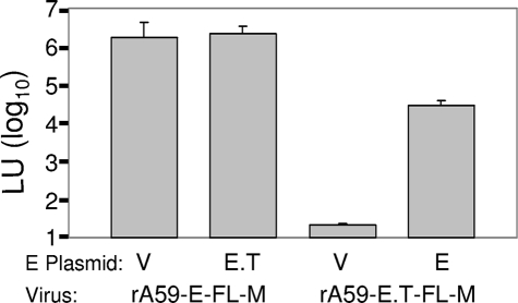
The coronavirus assembly process encloses a ribonucleoprotein genome into vesicles containing the lipid-embedded proteins S (spike), E (envelope), and M (membrane). This process depends on interactions with membranes that may involve palmitoylation, a common posttranslational lipidation of cysteine residues. To determine whether specific palmitoylations influence coronavirus assembly, we introduced plasmid DNAs encoding mouse hepatitis coronavirus (MHV) S, E, M, and N (nucleocapsid) into 293T cells and found that virus-like particles (VLPs) were robustly assembled and secreted into culture medium. Palmitate adducts predicted on cysteines 40, 44, and 47 of the 83-residue E protein were then evaluated by constructing mutant cDNAs with alanine or glycine codon substitutions at one or more of these positions. Triple-substituted proteins (E.Ts) lacked palmitate adducts. Both native E and E.T proteins localized at identical perinuclear locations, and both copurified with M proteins, but E.T was entirely incompetent for VLP production. In the presence of the E.T proteins, the M protein subunits accumulated into detergent-insoluble complexes that failed to secrete from cells, while native E proteins mobilized M into detergent-soluble secreted forms. Many of these observations were corroborated in the context of natural MHV infections, with native E, but not E.T, complementing debilitated recombinant MHVs lacking E. Our findings suggest that palmitoylations are essential for E to act as a vesicle morphogenetic protein and further argue that palmitoylated E proteins operate by allowing the primary coronavirus assembly subunits to assume configurations that can mobilize into secreted lipid vesicles and virions.


Figures

FIG. 1.
FIG. 1.
Features of coronavirus E proteins. The cysteine-rich region of the MHV E protein (residues 38 to 52) is depicted in a helical wheel format. Hydrophobic residues of the helical wheel are identified with large encircled shading, and palmitates extending from cysteines 40, 44, and 47 are depicted with corrugated lines. Cysteines 40, 44, and 47 are denoted with asterisks in the primary sequence. Residues 38 to 52 are delineated in the context of the entire E amino acid sequence and compared with E sequences for groups 1 (TGEV) and 3 (IBV). The membrane-intercalating portions of each E protein are highlighted. A predicted membrane organization for the MHV E protein is illustrated at the bottom, in the context of a budding virion, with the outside of the virion at left and the inside at right. See the text for additional details.
FIG. 2.
FIG. 2.
Plasmid DNA-encoded VLP production. Parallel cultures of 106 293T cells were transfected with 1-μg amounts of pCAGGS plasmids encoding the indicated MHV (strain A59) proteins, as described in Materials and Methods. After 2 days, medium was collected and cells were dissolved in a lysis buffer containing NP-40 and deoxycholate, while VLPs in clarified medium was pelleted by ultracentrifugation. Viral proteins associated with the cell lysates, and VLPs were detected after their separation by SDS-PAGE and immunoblotting with antibodies specific for S, N, M, and E. Sunc, uncleaved ∼180-kDa S protein precursor. S2, C-terminal S cleavage product. The positions of molecular mass markers are designated in kilodaltons at the right of each immunoblot panel.
FIG. 3.
FIG. 3.
Evaluation of HA-tagged E proteins in VLP assays. 293T cells were transfected with pCAGGS plasmids encoding the indicated proteins. EHA encodes E with an appended C-terminal 9-amino-acid epitope. After 2 days, viral S, N, and M proteins were detected in cell lysate and VLP fractions as described in the legend for Fig. 2. The positions of molecular mass markers are designated in kilodaltons. Sunc, uncleaved ∼180-kDa S protein precursor. S2, C-terminal S cleavage product.
FIG. 4.
FIG. 4.
Evaluation of mutant E proteins in VLP assays. 293T cells were transfected with pCAGGS plasmids encoding S, N, M, and the indicated E forms. E.S is E with the C40A substitution, E.D is E with the C40A/C44G substitution, and E.T is E with the C40A/C44G/C47G substitution. At 2 days posttransfection/infection, cells were dissolved and secreted VLPs or virions were pelleted by ultracentrifugation. S, N, and M proteins in cell lysates and secreted VLPs or virions were detected by Western immunoblotting to generate the bands in lanes 1 to 5. In parallel, a 293T cell culture identical to that described above was transfected with pcDNA3-CEACAM encoding the MHV receptor and infected 18 h later with authentic MHV strain A59 at a multiplicity of infection of 1. Proteins in cell lysates and secreted virions were then evaluated by Western immunoblotting to generate the bands in lane 6. In this assay, lysate-associated proteins were derived from 1/50 of each culture, whereas VLP and virion proteins were derived from 1/5 of each culture.
FIG. 5.
FIG. 5.
Effect of hydroxylamine on E protein electrophoretic mobilities. 17Cl1 cells were lipofected with pCAGGS-EHA or E.THA and dissolved 24 h later in nonionic detergent-containing lysis buffer. E proteins in clarified cell lysates were then immunoprecipitated with anti-HA antibodies and dissolved in SDS solubilizer. Aliquots of the EHA- and E.THA-containing samples were incubated either alone (−) or together (+) with 0.5 M hydroxylamine for 15 min, subjected to electrophoresis on 14% polyacrylamide gels, and then detected by immunoblotting using anti-HA antibodies.
FIG. 6.
FIG. 6.
Subcellular localization of EHA and E.THA proteins. 17Cl1 cells were lipofected with pCAGGS-E plasmids and subsequently infected with MHV A59. At 8 hpi, cells were fixed, permeabilized, and stained with FITC-conjugated anti-HA antibodies (green), with mouse anti-N antibodies, and with secondary Alexa 658 (red)-conjugated anti-mouse immunoglobulin antibodies. In a separate assay, E-transfected cells were stained with anti-HA (green) and anti-Golgin-97 and then with Alexa 658 (red)-conjugated antibodies. Images were captured by confocal microscopy.
FIG. 7.
FIG. 7.
Copurification of E and M proteins. 293T cells were transfected with plasmids encoding the indicated viral proteins. Two days later, cells were collected into lysis buffer and separated into insoluble (Ins) and soluble (Sol) fractions, as described in Materials and Methods. M and E proteins associated with the subcellular fractions were detected by immunoblotting with anti-M and anti-HA antibodies. Positions of the 29- and 20-kDa molecular mass markers are indicated at the right side of the immunoblot images.
FIG. 8.
FIG. 8.
Mobilization of M proteins from insoluble complexes by increasing E levels. 293T cells (106) were transfected with 1-μg amounts of pCAGGS plasmids encoding S, M, N, and either E or E.T. Where indicated, the input doses of E and E.T plasmids were increased 10-fold (10×), to 10 μg. After 2 days, cells were collected into lysis buffer and fractionated into insoluble (Ins) and soluble (Sol) material. VLPs were pelleted from medium by ultracentrifugation. Samples representing equivalent proportions of each culture were then subjected to immunoblotting to detect the M and E proteins. Magg, SDS-insoluble M protein aggregates.
FIG. 9.
FIG. 9.
Complementation of rA59-Eko-FL-M by plasmid-encoded E proteins. As schematically depicted in panel A, 17Cl1 cells were lipofected with the indicated pCAGGS-E constructs and infected 6 h later with rA59-Eko-FL-M (0.004 PFU/cell). After 18 h, medium was collected from producer cells, clarified, and used to inoculate 17Cl1 target cells. At 6 hpi, target cells were lysed. Luciferase light units (LU) in target cell lysates are plotted in panel B. Each data point represents the average luciferase levels from three parallel lipofection infection assays. V, data obtained from empty pCAGGS vector-transfected cultures. Uninf, data obtained from uninfected control cultures. Panel C depicts E protein-specific signals obtained after Western immunoblot analysis of the indicated producer cell lysates that were collected at 18 hpi. Error bars indicate standard deviations.
FIG. 10.
FIG. 10.
Complementation of rA59-E- and E.T-FL-M by plasmid-encoded E proteins. Complementation assays were performed as described in the legend for Fig. 9, with rA59-Eko replaced by the indicated recombinant viruses. Luciferase (LU) data points represent the average of the values obtained from three parallel complementation assays. V, data obtained from empty pCAGGS vector-transfected cultures.
FIG. 11.
FIG. 11.
Augmentation of rA59-E-FL-M assembly/secretion by plasmid-encoded E proteins. 17Cl1 producer cells were transfected with pCAGGS-E plasmids as indicated. Six hours later, the cells were infected with rA59-E-FL-M virus and, at 12 hpi, media were collected and producer cells were lysed for determination of luciferase accumulations. The medium was then clarified and used to infect 17Cl1 target cells, which were lysed at 6 hpi and evaluated for luciferase accumulations using a plate-reading luminometer. Error bars indicate standard deviations. V, data obtained from empty pCAGGS vector-transfected cultures.

References

    1. Bauer, M., and L. Pelkmans. 2006. A new paradigm for membrane-organizing and -shaping scaffolds. FEBS Lett. 5805559-5564. - PubMed
    1. Bos, E. C., W. Luytjes, H. V. van der Meulen, H. K. Koerten, and W. J. Spaan. 1996. The production of recombinant infectious DI-particles of a murine coronavirus in the absence of helper virus. Virology 21852-60. - PMC - PubMed
    1. Corse, E., and C. E. Machamer. 2002. The cytoplasmic tail of infectious bronchitis virus E protein directs Golgi targeting. J. Virol. 761273-1284. - PMC - PubMed
    1. Corse, E., and C. E. Machamer. 2003. The cytoplasmic tails of infectious bronchitis virus E and M proteins mediate their interaction. Virology 31225-34. - PMC - PubMed
    1. Curtis, K. M., B. Yount, and R. S. Baric. 2002. Heterologous gene expression from transmissible gastroenteritis virus replicon particles. J. Virol. 761422-1434. - PMC - PubMed

LinkOut - more resources