. 2008 Mar;82(6):2821-35.
doi: 10.1128/JVI.01821-07. Epub 2008 Jan 9.

Egress of light particles among filopodia on the surface of Varicella-Zoster virus-infected cells

Affiliations

Egress of light particles among filopodia on the surface of Varicella-Zoster virus-infected cells

John E Carpenter et al. J Virol. 2008 Mar.

Abstract

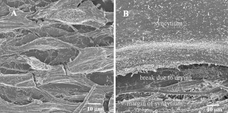
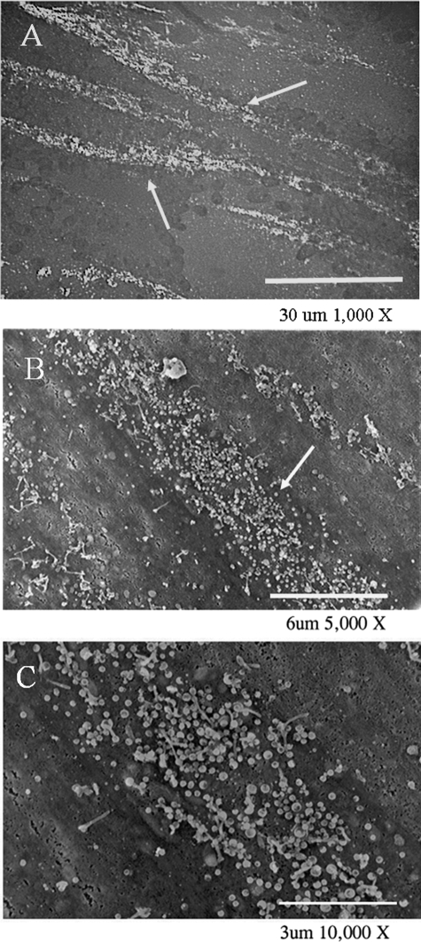
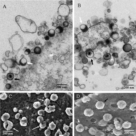
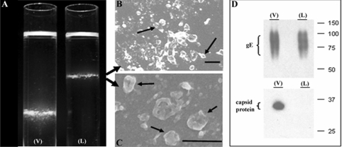
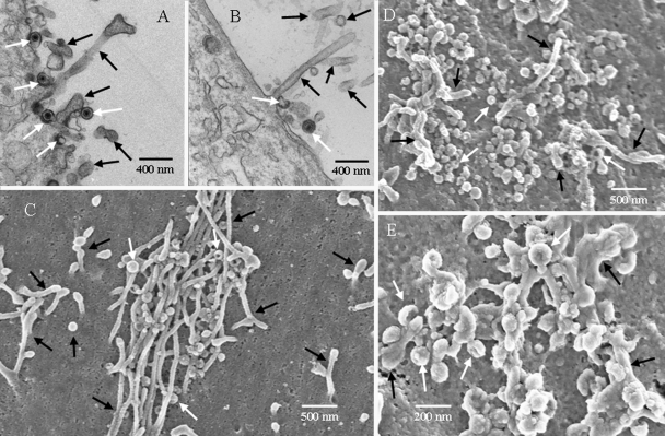
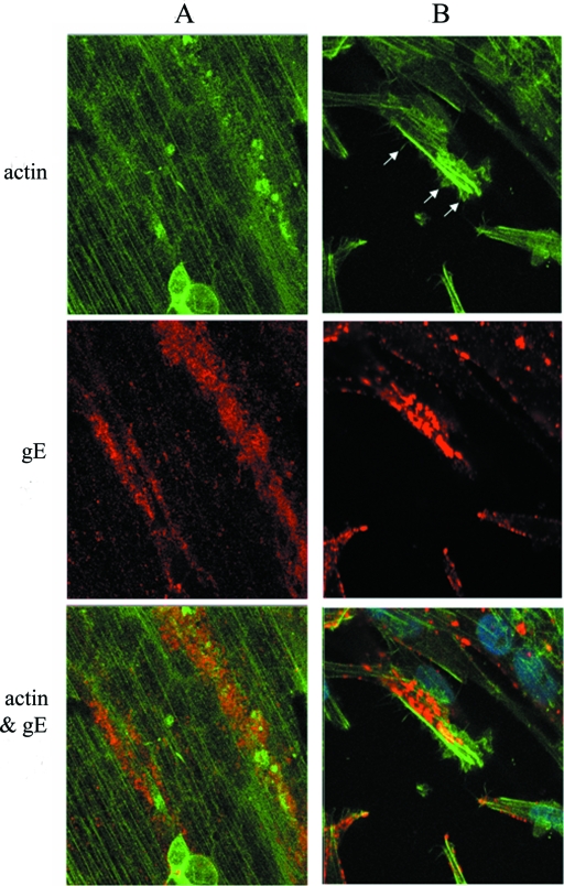
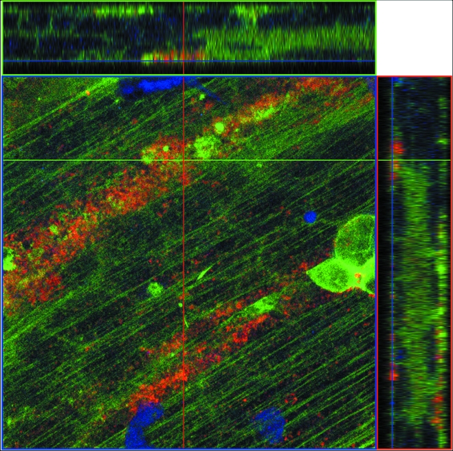
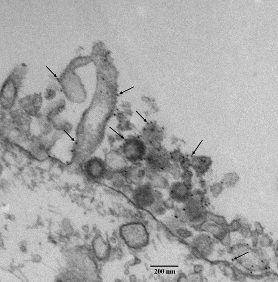
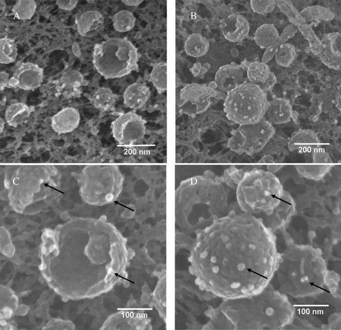
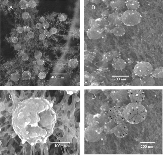
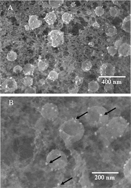
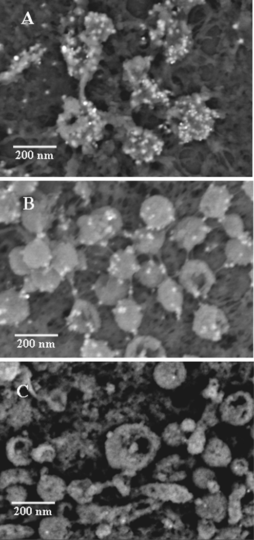
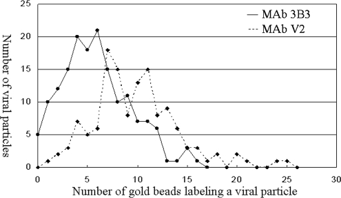
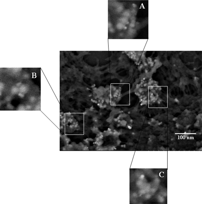
Varicella-zoster virus (VZV) is renowned for its very low titer when grown in cultured cells. There remains no single explanation for the low infectivity. In this study, viral particles on the surfaces of infected cells were examined by several imaging technologies. Few surface particles were detected at 48 h postinfection (hpi), but numerous particles were observed at 72 and 96 hpi. At 72 hpi, 75% of the particles resembled light (L) particles, i.e., envelopes without capsids. By 96 hpi, 85% of all particles resembled L particles. Subsequently, the envelopes of complete virions and L particles were investigated to determine their glycoprotein constituents. Glycoproteins gE, gI, and gB were detected in the envelopes of both types of particles in similar numbers; i.e., there appeared to be no difference in the glycoprotein content of the L particles. The viral particles emerged onto the cell surface amid actin-based filopodia, which were present in abundance within viral highways. Viral particles were easily detected at the base of and along the exterior surfaces of the filopodia. VZV particles were not detected within filopodia. In short, these results demonstrate that VZV infection of cultured cells produces a larger proportion of aberrant coreless particles than has been seen with any other previously examined alphaherpesvirus. Further, these results suggested a major disassociation between capsid formation and envelopment as an explanation for the invariably low VZV titer in cultured cells.


Figures

FIG. 1.
FIG. 1.
SEM images of uninfected and VZV-infected cells. (A) Uninfected monolayer with approximately 10 MeWo cells; (B) infected MeWo cells that have fused into a syncytium. Note that the drying process for SEM imaging leads to cell shrinkage and occasional breaks in the monolayer. In panel B, the margin of syncytium is visible near bottom of the image; above it is a wide break in the syncytial surface due to drying. The samples were prepared for viewing using protocol 2 (see Materials and Methods).
FIG. 2.
FIG. 2.
Typical patterns of VZV viral highways. (A) Epifluorescent microscopic image of two major highways (white arrows) and lesser highways immunostained by mouse MAb 3B3; (B and C) SEM images of viral highways (white arrows) at increasingly high magnifications, to document their width and the relative number of particles. Highways formed only in areas of the monolayer that were already fused, as evidenced by the lack of any detectable individual cells. The SEM samples were prepared for viewing using protocol 1 (see Materials and Methods).
FIG. 3.
FIG. 3.
Comparison of VZV particles by TEM (A and B) and SEM (C and D.) In all panels, black arrows indicate complete viruses and white arrows indicate aberrant particles; not all particles are indicated. (A) Seven VZV particles were visible, of which one was a complete virus. Most were envelopes without capsids. (B) Seven particles were visible, of which two contained capsids. Many particles in the TEM images were smaller than a full particle because the section was taken off center. (C) Sixteen particles were visible; the majority were aberrant. (D) Thirteen viral particles were visible, of which half were aberrant. The SEM samples were prepared for viewing using protocol 2 (see Materials and Methods).
FIG. 4.
FIG. 4.
Density gradient sedimentation of VZV particles. (A) Photograph of the bands after second sedimentation in 16- by 102-mm Beckman tubes; V, lower viral band; L, upper L-particle band. (B and C) Negative-staining SEM images of the upper band from the L tube, showing aberrant particles. (D) Immunoblot of bands from both tubes with anti-gE antibody and anticapsid protein antibody. Note the absence of detectable VZV capsid protein in the upper (L) band. Scale bars: panel B, 200 nm; panel C, 400 nm.
FIG. 5.
FIG. 5.
Microstructural composition of the viral highways. The viral highways included complete and aberrant viral particles (white arrows) and cellular projections (black arrows). The images show that the long cellular projections were 70 to 100 nm in diameter. (A and B) TEM images; (C to E) SEM images. (A) Five viral particles and several projections; (B) one elongated projection surrounded by portions of other projections; (C) viral highway covered by cellular projections with viral particles in between; (D) viral highway with predominantly viral particles and a few projections; (E) viral highway with projections that were closely intermingled with viral particles. Several viral particles were attached to the projections. The SEM samples were prepared for viewing using protocol 2 (see Materials and Methods).
FIG. 6.
FIG. 6.
Identification of F-actin in the viral highway. Infected monolayers were stained with phalloidin (green) and anti-gE MAb (red) before imaging by confocal microscopy. Two images were obtained in different areas of the infected monolayer (A and B). (A) VZV gE and F-actin colocalized along a viral highway oriented along the terminal web stained by F-actin. Multinucleated syncytia are visible in the infected monolayer. (B) VZV gE and F-actin staining of infected MeWo cells showing cellular projections (white arrows) stained for F-actin. In order to best show the projections, a cell that was not adjacent to another cell was selected.
FIG. 7.
FIG. 7.
Colocalization of F-actin and VZV gE on the cellular surface. Cross-sectional views of actin (green) and gE (red) were visualized in a combined image by confocal microscopy. The central image is in the xy plane. The top image is a cross-section in the xz plane along the green line. The right-side image is a cross section in the yz plane along the red line. Eighteen overlapping 1-μm slices were taken in the z direction. The two cross-sectional images revealed that the gE staining was localized on the cellular surface.
FIG. 8.
FIG. 8.
Detection of VZV gE in the viral highway by electron microscopy. An infected monolayer was labeled with mouse MAb 3B3 and 10-nm gold beads. The attachment of gold beads to viral particles and filopodia was documented by TEM imaging. Note the absence of a viral particle within the elongated cellular projection.
FIG. 9.
FIG. 9.
Detection of VZV gE on viral particles by electron microscopy. Samples were immunolabeled with MAb 3B3 and silver-enhanced ultrasmall gold beads and visualized by SEM. Arrows indicate gold beads. Not all beads are indicated. (A) Twelve viral particles with four or five beads each. (B) Thirteen particles with four or five beads each; the large complete particle in the middle has fourteen beads. (C) Enlargement of panel A with beads on three particles. (D) Enlargement of the large particle in panel B to further delineate the gold beads labeling gE. The immunolabeled samples in this figure and subsequent figures were prepared for viewing using protocol 1 (see Materials and Methods).
FIG. 10.
FIG. 10.
Detection of VZV gI on viruses by electron microscopy. Samples were immunolabeled with mouse MAb 6B5 and silver-enhanced ultrasmall gold beads and visualized with SEM. Arrows indicate gold beads. Not all beads are indicated. (A) Twenty-five particles, each with beads. (B) Fourteen particles with four or six beads each. (C) Enlargement of a single viral particle with ten beads surrounding a gap in the envelope. (D) Enlargement of panel B, to show that beads were often visualized around the circumference of an indentation in the envelope.
FIG. 11.
FIG. 11.
Detection of VZV gB on viruses by electron microscopy. Samples were immunolabeled with human MAb V1 and silver-enhanced ultrasmall gold beads and visualized by SEM. Arrows indicate gold beads (not all beads are indicated). (A) Twenty viral particles, all of which had beads. (B) Enlargement of several viral particles. Each particle contained three or four beads.
FIG. 12.
FIG. 12.
Effect of primary antibody and gold bead size on VZV gE as seen in backscatter images. (A) Imaging of human MAb V2 and ultrasmall gold beads, showing 10 viral particles with numerous gold beads. (B) Imaging of mouse MAb 3B3 with ultrasmall gold beads, showing 24 viral particles labeled by fewer gold beads than in panel A. (C) Imaging of mouse MAb 3B3 with 10-nm gold beads, showing 12 viral particles with fewer gold beads than in either panel A or B.
FIG. 13.
FIG. 13.
Number of VZV viral particles with a given number of gold beads. Two separate monolayers were immunolabeled, one with mouse MAb 3B3 (circles) and one with human MAb V2 (diamonds). After immunogold labeling, beads in SEM images were counted.
FIG. 14.
FIG. 14.
Multimeric structure of VZV gE. Infected cells were labeled with human MAb V2 and observed in high-resolution SEM images. Each panel showed envelope structures labeled by compact and diffuse signals of backscatter, indicating gE dimers. (A) Two gE dimers arranged in parallel; (B) two gE dimers arranged antiparallel; (C) multiple gE dimers arranged in a cluster.

References

    1. Bozzola, J. J., and L. D. Russell. 1992. Electron microscopy. Jones and Bartlett Publishers, Boston, MA.
    1. Cook, M. L., and J. G. Stevens. 1968. Labile coat: reason for non-infectious cell-free varicella-zoster virus in culture. J. Virol. 21458-1464. - PMC - PubMed
    1. Cook, M. L., and J. G. Stevens. 1970. Replication of varicella-zoster in cell culture: an ultrastructural study. J. Ultra. Res. 32334-350. - PubMed
    1. Favoreel, H. W., G. Van Minnebruggen, D. Adriaensen, and H. J. Nauwynck. 2005. Cytoskeletal rearrangements and cell extensions induced by the US3 kinase of an alphaherpesvirus are associated with enhanced spread. Proc. Natl. Acad. Sci. USA 1028990-8995. - PMC - PubMed
    1. Friedrichs, W. E., and C. Grose. 1986. Varicella-zoster virus p32/p36 complex is present in both the viral capsid and the nuclear matrix of the infected cell. J. Virol. 57155-164. - PMC - PubMed

Publication types

LinkOut - more resources