. 2008 Mar;82(6):3131-4.
doi: 10.1128/JVI.02266-07. Epub 2008 Jan 9.

Cysteines flanking the internal fusion peptide are required for the avian sarcoma/leukosis virus glycoprotein to mediate the lipid mixing stage of fusion with high efficiency

Affiliations

Cysteines flanking the internal fusion peptide are required for the avian sarcoma/leukosis virus glycoprotein to mediate the lipid mixing stage of fusion with high efficiency

Sue E Delos et al. J Virol. 2008 Mar.

Abstract

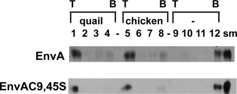
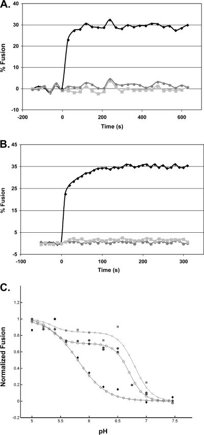
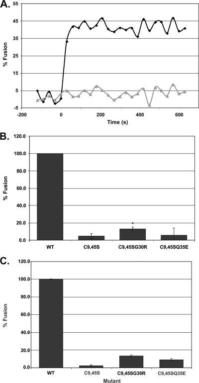
We previously showed that the cysteines flanking the internal fusion peptide of the avian sarcoma/leukosis virus subtype A (ASLV-A) Env (EnvA) are important for infectivity and cell-cell fusion. Here we define the stage of fusion at which the cysteines are required. The flanking cysteines are dispensable for receptor-triggered membrane association but are required for the lipid mixing step of fusion, which, interestingly, displays a high pH onset and a biphasic profile. Second-site mutations that partially restore infection partially restore lipid mixing. These findings indicate that the cysteines flanking the internal fusion peptide of EnvA (and perhaps by analogy Ebola virus glycoprotein) are important for the foldback stage of the conformational changes that lead to membrane merger.


Figures

FIG. 1.
FIG. 1.
Receptor-triggered membrane association of EnvA and EnvAC9,45S. Virus-liposome association assays were modified from the work of Netter et al. (15). Briefly, 40 μl of virus was mixed with sTva (final concentration, 1 μM) and allowed to associate for 30 min on ice. After addition of 50 μl of liposomes (a 1:1:1:1.5 mixture of phosphatidylcholine, PE, sphingomyelin, and cholesterol; extruded through 100-nm-pore-size filters) and additional HM buffer (20 mM HEPES, 20 mM morpholineethanesulfonic acid, 130 mM NaCl [pH 7.5]) to bring the final volume to 100 μl, the samples were incubated at 37°C for 30 min and then returned to ice. The virus-liposome mixture was then diluted 1:1 with 80% (wt/vol) sucrose in HM buffer, transferred to a 700-μl centrifuge tube, and overlaid first with 400 μl of 25% sucrose and then with 50 μl of 5% sucrose. Samples were centrifuged at 150,000 × g in a 55 Ti rotor for 2 h. Four fractions, of 100, 150, and 150 μl and the remainder (approximately 200 μl), were collected from the top of the gradient. Proteins were resolved by sodium dodecyl sulfate-polyacrylamide gel electrophoresis, transferred to nitrocellulose filters, probed with an anti-A tail antibody (7), and visualized with a horseradish peroxidase-conjugated anti-rabbit antibody. Either the quail or the chicken isoform of sTva or no receptor (-) was used to trigger membrane binding, as indicated. T, top of the gradient; B, bottom of the gradient; sm, a portion (1/10) of the starting sample used for the given triggering experiment.
FIG. 2.
FIG. 2.
Characterization of lipid mixing induced by ASLV and VSV-EnvA. (A and B) Virus from infected cell culture medium was concentrated in 300,000-molecular-weight-cutoff Vivaspin columns and purified by banding on a step gradient of 25% and 60% sucrose (wt/vol). The visible band was collected, pelleted and concentrated by centrifuging through 20% sucrose, and resuspended in 150 μl HM buffer (20 mM HEPES, 20 mM morpholineethanesulfonic acid, 130 mM NaCl [pH 7.5]) overnight on ice at 4°C. To assay lipid mixing, 25 μg ASLV-A (A) or VSV-EnvA (B) was first incubated with sTva (solid black diamonds and solid light grey squares) or without sTva (solid dark grey circles) at pH 7.5 for 30 min at 4°C; liposomes (a 1:1:1:1.5:0.045:0.11 mixture of phosphatidylcholine, PE, sphingomyelin, cholesterol, Rh-PE, and NBD-PE) prepared by extrusion through 50-nm-pore-size filters (6) were added; and the mixture was warmed to 37°C for 15 min to trigger target membrane binding, after which baseline fluorescence at pH 7.5 was recorded for 5 min at 37°C. At time zero, the samples were adjusted to pH 5 and incubated at 37°C, and fluorescence (excitation at 460 nm; emission at 540 nm) was measured for 10 min. Where indicated, R99 was added to a final concentration of 100 μg/ml prior to acidification (solid light grey squares). Maximum possible NBD fluorescence was determined by adding NP-40 to a final concentration of 1% and measuring fluorescence at 37°C for 15 min. Percent fusion was calculated as (FpHF0)/(FTF0) × 100, where F0 is the baseline fluorescence (pH 7.5), FpH is the averaged fluorescence at the plateau at pH 5.0, and FT is the fluorescence at an infinite dilution (after disruption of the membranes with 1% NP-40). The data from triplicate samples were averaged. Results of a representative experiment are shown. Each experiment was repeated two or more times. (C) pH dependence of lipid mixing. Samples were treated as for panels A and B except that the pH was adjusted as indicated on the x axis. The results from triplicate samples of individual experiments were averaged, the value at pH 5 was set to 1, and fusion at each individual pH was reported as a fraction of the pH 5 value. The data from two independent ASLV-A experiments (solid dark grey circles), one VSV-EnvA experiment (solid light grey squares), and one influenza X:31 virus (Charles River Laboratories) experiment are plotted. The data for each virus were then fitted by a nonlinear least-squares method using the MatLab curve-fitting toolbox and the equation % fusion = c{1 − tanh[d(pH − pH0)]}, where pH0 is the pH at the inflection point of the curve, c is one-half the height between the initial and the maximal fusion for the curve, and the product(c·d) is the slope of the curve at the inflection point. The data from two independent ASLV experiments (filled circles) and one VSV-EnvA experiment (shaded squares) are plotted. For comparison, the pH dependence of influenza virus X:31 (Charles River Laboratories) was determined (filled diamonds). The fit curves are plotted using the corresponding open circles, X's, and open diamonds, respectively.
FIG. 3.
FIG. 3.
Characterization of lipid mixing induced by VSV-EnvAC9,45S and two second-site revertants. (A) Lipid mixing induced by VSV-EnvA (diamonds) and VSV-EnvAC9,45S (triangles). (B and C) Extent of lipid mixing for VSV-EnvA (wild type [WT]), VSV-EnvAC9,45S, VSV-EnvAC9,45SG30R, and VSV-EnvAC9,45SQ35E. Virus samples were prepared and lipid mixing assessed as described for Fig. 2, except that the relative amount of each virus used per assay was normalized for the relative incorporation of the various EnvA's (ranges, 0.63 to 1.42 for EnvAC9,45S, 0.63 to 0.86 for EnvAC9,45SG30R, and 0.71 to 1.02 for EnvAC9,45SQ35E, relative to wild-type EnvA) and the fusion observed for wild-type EnvA in the presence of R99 was used as pH0. Data in panel B are averages from four experiments, each carried out in triplicate. t tests were performed using the “two sample assuming unequal variances” function in Microsoft Excel [*, P(Tt) = 0.0037]. Data in panel C are averages from duplicate samples from a single experiment. In panels B and C, FpH was calculated as for Fig. 2C.

References

    1. Armstrong, R. T., A. S. Kushnir, and J. M. White. 2000. The transmembrane domain of influenza hemagglutinin exhibits a stringent length requirement to support the hemifusion to fusion transition. J. Cell Biol. 151425-437. - PMC - PubMed
    1. Baker, K. A., R. E. Dutch, R. A. Lamb, and T. S. Jardetzky. 1999. Structural basis for paramyxovirus-mediated membrane fusion. Mol. Cell 3309-319. - PubMed
    1. Barnard, R. J., S. Narayan, G. Dornadula, M. D. Miller, and J. A. Young. 2004. Low pH is required for avian sarcoma and leukosis virus Env-dependent viral penetration into the cytosol and not for viral uncoating. J. Virol. 7810433-10441. - PMC - PubMed
    1. Delos, S. E., J. M. Gilbert, and J. M. White. 2000. The central proline of an internal viral fusion peptide serves two important roles. J. Virol. 741686-1693. - PMC - PubMed
    1. Delos, S. E., and J. M. White. 2000. Critical role for the cysteines flanking the internal fusion peptide of avian sarcoma/leukosis virus envelope glycoprotein. J. Virol. 749738-9741. - PMC - PubMed

Publication types

LinkOut - more resources