. 2008 Mar;146(3):1165-81.
doi: 10.1104/pp.107.114751. Epub 2008 Jan 9.

SEUSS and AINTEGUMENTA mediate patterning and ovule initiation during gynoecium medial domain development

Affiliations

SEUSS and AINTEGUMENTA mediate patterning and ovule initiation during gynoecium medial domain development

Sridevi Azhakanandam et al. Plant Physiol. 2008 Mar.

Abstract

The Arabidopsis (Arabidopsis thaliana) gynoecium, the female floral reproductive structure, requires the action of genes that specify positional identities during its development to generate an organ competent for seed development and dispersal. Early in gynoecial development, patterning events divide the primordium into distinct domains that will give rise to specific tissues and organs. The medial domain of the gynoecium gives rise to the ovules, and several other structures critical for reproductive competence. Here we report a synergistic genetic interaction between seuss and aintegumenta mutants resulting in a complete loss of ovule initiation and a reduction of the structures derived from the medial domain. We show that patterning events are disrupted early in the development of the seuss aintegumenta gynoecia and we identify PHABULOSA (PHB), REVOLUTA, and CRABS CLAW (CRC) as potential downstream targets of SEUSS (SEU) and AINTEGUMENTA (ANT) regulation. Our genetic data suggest that SEU additionally functions in pathways that are partially redundant and parallel to PHB, CRC, and ANT. Thus, SEU and ANT are part of a complex and robust molecular system that coordinates patterning cues and cellular proliferation along the three positional axes of the developing gynoecium.


Figures
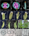
Figure 1.
Figure 1.
seu ant double mutants display enhanced vegetative, floral, and gynoecial phenotypes. A to C, False-colored photomicrographs. D to K, Photomicrographs of floral/rosette morphology; some sepals and petals have been removed from front half of flowers to allow viewing of gynoecium. L and M, Alcian-blue-stained gynoecial cross sections; N and O, Longitudinal optical section of cleared gynoecia (Nomarski optics). Scale bars: D to J, 1 mm; K, 10 mm; L to O, 0.1 mm. Asterisks (*) in M and O indicate expected location of ovules. A and B, Gynoecial cross sections at level of ovary from stage 8 (A) and stage 12 (B) flowers. Medial and lateral domains are shown subdivided into adaxial (inner) and abaxial (outer) portions by dotted oval. Two medial ridges (mr) arise on adaxial portions of medial domain (purple/magenta) at stage 8 and give rise to ovules (ov) and septum (s) in mature gynoecium. The abaxial replum (abr) forms from the abaxial portion of the medial domain (orange). The lateral domains gives rise to carpel valves (cv). C, Side view of mature gynoecium indicates patterning elements along apical basal axis: stigma (stg), style (sty), and ovary (ovy). D, Wild-type Col-0; E, ant-3 mutant; F, ant-1 mutant, petals narrower than wild type (arrowhead); G, seu-3 mutant; H, seu-3 ant-3 double mutant displays enhanced carpel splitting at apex (arrowhead); I and J, seu-3 ant-1 double mutant displays filamentous petals (fp) and very reduced stamens (st); gynoecium (g) is split open and does not display ovules; medial domain displays small amounts of stigmatic (stg) and stylar tissue. K, Rosette phenotypes of Col-0 (left) and seu-3 ant-1 (right). L, In Col-0 gynoecium transmitting tract (tt) stains blue and ovules (ov) are indicated. M, Transmitting tract stains blue in the seu-3 ant-1 double mutant whereas ovules are missing. N, Developing ovules (ov) are observed attached to septum (s) in the seu-3 ant-3 double mutant. O, In the seu-3 ant-1 double mutant, no ovules grow out from septum (s).
Figure 2.
Figure 2.
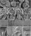
Early floral and ovule defects in seu ant double mutants. A to O, SEM micrographs. Numbers refer to stages of floral development. P to U, Nomarski contrast images of optically cleared tissue. Scale bars in A to C and H to O, 100 microns; bars in D to G and R to U, 50 microns; bars in P and Q, 500 microns. A, Col-0 inflorescence. Floral meristems display four sepals (se). Inflorescence meristem (ifm). B and C, seu-3 ant-1 floral meristems display three narrow sepals. Locations of missing sepals indicated by asterisks (*). D to G, Stage-6 gynoecia of indicated genotypes. Black line indicates plane of medial domain. Arrowheads indicate medial ridges. Medial ridges appear reduced in G. H, Stage-11 Col-0 gynoecia. I, Stage-11 seu-1 ant-3 gynoecium with enhanced apical splitting. J, Mature Col-0 gynoecium. K, Mature seu-3 ant-1 flower. Gynoecium split at apex. Lateral carpel horns (lh) and medial domains (md) marked. Stamens (st) and sepals (se) reduced in size. L), Adaxial Col-0 leaf surface. M, Abaxial Col-0 leaf surface. N, Adaxial seu-3 leaf surface. O, Adaxial seu-3 ant-1 double-mutant leaf surface. Cells appear intermediate between adaxial and abaxial identities. P, Apex of seu-3 ant-3 gynoecium. Vascular bundle in medial domain branches into stylar vascular array (arrowhead) similar to wild type. Lateral vascular bundle reaches apex (arrow). Q, Basal region of seu-3 ant-1 gynoecium. Medial bundle terminates prematurely (arrowhead). Lateral bundle (arrow) reaches apex (not shown). R to U, Mature ovules of indicated genotypes. Arrowhead indicates central cell (R and S) or partially developed female gametophyte (T). The asterisk (*) in U indicates expected location of female gametophyte.
Figure 3.
Figure 3.
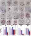
Adaxial fate determinants PHB and REV in seu ant gynoecia. In situ hybridization with PHB antisense (A–M) or REV antisense (N–T) probes. A, D, F, G, J, K, L, and M, Floral longitudinal sections. B, C, E, H, I, and N to T, Floral cross sections (extent of gynoecia encircled by black dashed ovals). All scale bars are 0.1 mm. Numbers refer to floral stages. A, PHB expression detected in central core of stage-3 Col-0 flower. B and C, PHB expression detected in adaxial core of gynoecial (arrowhead) and stamen (arrow) anlagen/primordia. D, PHB expression in gynoecium (arrowhead). E, PHB expression in adaxial valve domains of gynoecium (arrowheads). F, PHB expression in placenta and ovule primordia (arrowhead). G, PHB expression detected in central core of ant-1 floral meristem. H and I, PHB expression is reduced or not detected in ant-1 gynoecia (arrowheads). Expression in stamens is detected (arrows). J, PHB expression in placenta and ovule primordia (arrowhead) is reduced in ant-1. K, PHB expression is detected in stage-3 floral meristems of seu ant mutant. L and M, PHB expression is reduced in gynoecium (arrowheads). N to P, REV expression detected in the central portion of Col-0 gynoecium; stages 6 to 9. Q and R, REV expression detected in central portion of seu ant gynoecia (arrowheads). S and T, REV expression in gynoecia (stages 8 and 9) appears slightly reduced relative to wild type (compare S with O and T with P). U and V, qRT-PCR results. Relative expression levels for indicated genotypes from inflorescence apex (U) and stages 8 through 10 gynoecia (V). Statistically significant difference versus wild type; single asterisk (*), P < 0.05; triple asterisk (***), P < 0.001.
Figure 4.
Figure 4.
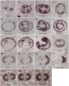
Abaxial fate determinants YAB1/FIL and CRC in seu ant gynoecia. In situ hybridization gynoecial cross sections. Probes in A to K, CRC; L, ANT; M to S, YAB1/FIL. A to L are at same scale. Scale bar in A, M to N, and Q to S, 0.1 mm; bar in O to P, 0.01 mm. A, Stage-6 wild-type section. Expression of CRC detected in two lateral valve domains (arrowheads). B, Expression of CRC at stage 7 observed in abaxial-most epidermal cells (arrow) and two internal stripes (arrowheads). C, Expression at stage 8 in abaxial epidermal cells, in four internal domains (arrowheads) and in adaxial valve epidermis (asterisk). D, Apical cross section, stage 8. CRC detected in valve domains (v) and more strongly in abaxial margin domains (abm), but is not detected in adaxial margin domains/medial ridge (mr). E to G, Serial gynoecial cross sections, each 24 microns apart. E, Basal section, seu ant gynoecium. CRC expression detected in abaxial epidermis, but no expression detected in internal domains. F, Midgynoecial section. CRC expression sometimes detected in internal domains, but domains are smaller and less consistently detected. Expression is detected in adaxial valve epidermis (asterisk). The lowermost of two medial ridges in this section appears reduced in size. G, Apical cross section. Ectopic expression of CRC detected in medial ridge (mr; compare to D). H, Midgynoecial section; only one of two medial ridges (mr) is detected. Ectopic epidermal (ec*) expression of CRC detected where medial ridge cells are expected. I to K, Serial gynoecial cross sections, each 24 microns apart. I, Basal cross section displays limited internal expression domains (arrowhead) while maintaining abaxial epidermal expression (arrow). J, Midgynoecial cross section. Limited expression in internal domains. K, Apical cross section. CRC ectopically detected in medial ridges. L, ANT expression in apical medial ridge (stage 8). M and N, Stage-9 cross sections of indicated genotypes. YAB1/FIL expression detected in abaxial valve mesophyl cells. Fingers of weaker expression (asterisk) extend into portions of medial domain that subtend ovules. These fingers are less consistently observed in the seu ant mutant (N). O and P, Stage-7 apical cross sections. YAB1/FIL expression is not detected in medial ridge (mr). Q, R, and S, Three serial cross sections (stage 8). Fingers of YAB1/FIL expression (asterisk) are often, but not always, detected subtending the sites of ovule initiation.
Figure 5.
Figure 5.
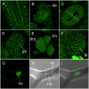
Expression of genomic SEUSS_GFP rescue construct. All images are from seu-1 mutant plants phenotypically rescued by the pSEU∷GFP:SEU fusion construct. A, pSEU∷GFP:SEU expression in root division zone is detected in nearly all cells in the nucleus (nucleoplasm), but is not detected or is significantly reduced in the nucleolus and cytoplasm. B, Expression in the stage-3 floral bud optical longitudinal section is detected in all whorls; sepal (se). C, A stage-8 gynoecial optical cross section. Expression is detected throughout the gynoecium. Medial plane is indicated by line. Apparent cytoplasmic florescence is due to a difficulty imaging deep tissue layers and likely represents background florescence, not bona fide GFP:SEU expression. D, Stage-8 gynoecium (gy), optical longitudinal section. E, Inflorescence meristem (ifm) and young floral meristems (fm). F, Young rosette leaf primordium (lp). G to I, Trichome cell. G, GFP image. Expression is seen in nucleus, but not nucleolus (no), nor cytoplasm. H, Nomarski contrast image showing nucleolus (no) and cytoplasm (cy). I, Merged G and H.
Figure 6.
Figure 6.
The seuss crabs claw double mutants display enhanced gynoecial defects. Photomicrographs of mature gynoecia of indicated genotypes. Scale bars are 1 mm for all panels. A, Landsberg erecta (Ler) reference ecotype. B, Gynoecium of the crabs claw (crc-1) allele is shorter and wider than wild type with mild splitting at apex (arrowhead). C, Gynoecium of seu-1 allele is slightly split at apex (arrowhead in inset). D, seu-1 crc-1 double-mutant gynoecium displays enhanced splitting of carpels, exposing ovules (arrowhead). Valve horns (vh) are also exhibited.
Figure 7.
Figure 7.
SEU and ANT function in parallel to PHB. A to F, Rosette phenotypes. G to L, Floral phenotypes of indicated genotypes. All plants grown at 16°C. Scale bars, A and B, 3 mm; C, 1.5 mm; D to K, 1 mm; and L, 0.5 mm. A, Wild-type Landsberg erecta (Ler). Primary bolt has been removed. B, phb-1d/+ plant. Rosette leaves display longer petiole (p) and smaller blade (b) than wild type. C, Rosette leaves of phb-1d/+ seu-3 ant-1 are narrower and more strongly radialized than phb-1d/+ plants. D, phb-1d homozygous mutant. Rosette leaves display limited laminar expansion (le) and examples of trumpet (tr) shaped leaves. E, Rosette leaves of phb-1d seu-3 display enhanced radialization and narrowing relative to phb-1d homozygote. F, Rosette leaves of phb-1d seu-3 ant-1 triple mutant are similar to phb-1d seu-3 double. G, Ler. H, ant-1 displays narrow petals (arrowhead). I, phb-1d/+. Petals are slightly narrower, particularly in basal portion (arrowhead). J, phb-1d/+ ant-1 displays filamentous petals (arrowhead). K, phb-1d/+ seu-3 also displays filamentous petals. L, phb-1d/+ seu-3 ant-1 is phenotypically similar to seu-3 ant-1 double and displays severely split gynoecium. Margin domains (md) completely lack ovule primordia.
Figure 8.
Figure 8.
A model for the action of SEU, LUG, ANT, and YAB1/FIL in ovule initiation and gynoecium medial domain development SEU physically interacts with LUG to form a transcriptional coregulator complex. In wild-type plants the SEU/LUG coregulator complex works together with ANT and YAB1/FIL to regulate PHB and REV expression and maintain adaxial identity within the developing gynoecium. In parallel to the maintenance of PHB expression, the SEU, LUG, ANT, and YAB1/FIL activities stimulate cell proliferation in the developing gynoecium. SEU, LUG, and ANT also engender a position-dependent regulation of CRC expression: supporting CRC expression in the internal expression domains of the gynoecium and repressing CRC expression at the gynoecial apex. These three functions support ovule initiation and medial domain development in wild-type gynoecia.

References

    1. Alvarez J, Smyth DR (1999) CRABS CLAW and SPATULA, two Arabidopsis genes that control carpel development in parallel with AGAMOUS. Development 126 2377–2386 - PubMed
    1. Alvarez J, Smyth DR (2002) CRABS CLAW and SPATULA genes regulate growth and pattern formation during gynoecium development in Arabidopsis thaliana. Int J Plant Sci 163 17–41
    1. Balanza V, Navarrete M, Trigueros M, Ferrandiz C (2006) Patterning the female side of Arabidopsis: the importance of hormones. J Exp Bot 57 3457–3469 - PubMed
    1. Berleth T, Jurgens G (1993) The role of monopteros in organizing the basal body region of the Arabidopsis embryo. Development 118 575–587
    1. Bowman JL, Baum SF, Eshed Y, Putterill J, Alvarez J (1999) Molecular genetics of gynoecium development in Arabidopsis. Curr Top Dev Biol 45 155–205 - PubMed

Publication types