. 2008 Mar;19(3):1113-24.
doi: 10.1091/mbc.e07-08-0742. Epub 2008 Jan 9.

The sequential activation of the yeast HOG and SLT2 pathways is required for cell survival to cell wall stress

Affiliations

The sequential activation of the yeast HOG and SLT2 pathways is required for cell survival to cell wall stress

Clara Bermejo et al. Mol Biol Cell. 2008 Mar.

Abstract

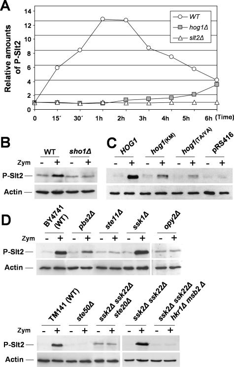
Yeast mitogen-activated protein kinase (MAPK) signaling pathways transduce external stimuli into cellular responses very precisely. The MAPKs Slt2/Mpk1 and Hog1 regulate transcriptional responses of adaptation to cell wall and osmotic stresses, respectively. Unexpectedly, we observe that the activation of a cell wall integrity (CWI) response to the cell wall damage caused by zymolyase (beta-1,3 glucanase) requires both the HOG and SLT2 pathways. Zymolyase activates both MAPKs and Slt2 activation depends on the Sho1 branch of the HOG pathway under these conditions. Moreover, adaptation to zymolyase requires essential components of the CWI pathway, namely the redundant MAPKKs Mkk1/Mkk2, the MAPKKK Bck1, and Pkc1, but it does not require upstream elements, including the sensors and the guanine nucleotide exchange factors of this pathway. In addition, the transcriptional activation of genes involved in adaptation to cell wall stress, like CRH1, depends on the transcriptional factor Rlm1 regulated by Slt2, but not on the transcription factors regulated by Hog1. Consistent with these findings, both MAPK pathways are essential for cell survival under these circumstances because mutant strains deficient in different components of both pathways are hypersensitive to zymolyase. Thus, a sequential activation of two MAPK pathways is required for cellular adaptation to cell wall damage.


Figures

Figure 1.
Figure 1.
CRH1 is induced under cell wall damage conditions. (A) The expression of CRH1 was examined after Congo Red (30 μg/ml) and zymolyase (0.4 U/ml) treatment in the WT strain (BY4741) carrying the pCRH1-LACZ plasmid. Cells were collected at different time points, and both CRH1 mRNA levels and β-galactosidase activity were determined, as described in Materials and Methods. Congo Red treatment corresponds to black bars (β-galactosidase activity) and squares (CRH1 mRNA). Zymolyase is represented by white bars (β-galactosidase activity) and squares (CRH1 mRNA). In both cases, the results are expressed as the ratio of treated versus untreated cells. Data represent means and SDs of three independent experiments. (B) BY4741 WT cells expressing Crh1-GFP were analyzed by flow cytometry after treatment with 0.4 U/ml zymolyase (gray line) or nontreated (black line) at the different times indicated. Dotted line corresponds to control BY4741 cells not expressing Crh1-GFP. (C) Localization of Crh1-GFP in WT cells (BY4741) growing exponentially in YPD and treated for 3 h with zymolyase (0.4 U/ml; right) or not treated (left).
Figure 2.
Figure 2.
Adaptation to zymolyase requires different elements of the Slt2 MAPK pathway but not the sensors or the GEFs of this pathway. (A) Time course of Slt2 activation in WT (BY4741) cells growing at 24°C to midlog phase and exposed to zymolyase (0.4 U/ml) at the indicated times. The protein load was monitored using a mouse anti-actin mAb. (B) Phosphorylation of Slt2 after 2 h of zymolyase treatment in slt2Δ, mkk1Δ mkk2Δ, bck1Δ, and pkc1Δ mutants. (C and D) Expression of CRH1-lacZ was determined in WT, slt2Δ, bck1Δ, rlm1Δ, swi4Δ, and swi6Δ strains (C) and WT, mid2Δ, wsc1Δ, wsc2Δ, wsc3Δ, wsc4Δ, and mtl1Δ strains (D), in the absence (□) and presence (3 h) of zymolyase 100T (0.4 U/ml; ■). (E) Slt2-phosphorylation by zymolyase in mid2Δ, wsc1Δ, and mid2Δ wsc1Δ double mutant (strain HAS17-3D) compared with corresponding WT strains, BY4741 and HAS17-3B. (F) Slt2 activation in the rom2Δ mutant compared with the WT (MCH-7B) in untreated cells or cells growing in the presence of Congo Red (30 μg/ml) or zymolyase (0.4 U/ml) for 2 h. (G) Phosphorylation of Slt2 in rom1Δ, rom2Δ, and tus1Δ mutants (BY4741 background) after 2 h of zymolyase treatment. Graphics in F and G represent quantification, by densitometric analysis, of the Phospho-Slt2 bands from Western blots shown above, normalized with respect to the actin bands.
Figure 3.
Figure 3.
CRH1 expression is regulated by the MAPK Slt2 or by Hog1 and Slt2 MAPKs simultaneously, depending on the nature of the cell wall stress. (A) Expression of CRH1-LacZ was studied in WT (BY4741) and slt2Δ, hog1Δ, kss1Δ, and fus3Δ mutant cells growing in presence of Congo Red (30 μg/ml; ■) or zymolyase (0.4 U/ml; □). β-Galactosidase activities are expressed as the ratio of Congo Red or zymolyase-treated cells versus untreated cells. Mean and SDs derived from three independent experiments. (B) Levels of Crh1-GFP were examined by Western blot in WT BY4741, slt2Δ, and hog1Δ untreated and treated (Congo Red or zymolyase) cells transformed with plasmid pJV89GL. The protein load was determined using an anti-actin antibody.
Figure 4.
Figure 4.
Hog1 is slightly activated by zymolyase and the presence of an active form of this MAPK is necessary for the induction of CRH1. (A) Effect of zymolyase in Hog1 activation. Exponentially growing WT cells were collected before and after different intervals of zymolyase treatment, and Hog1 activation was examined by immunoblotting total extracts with an anti-phospho-p38 antibody. The levels of phosphorylation of Hog1 by 0.4 M NaCl are also shown. The phosphorylation of Hog1 by zymolyase was dependent on the presence of the MAPKK Pbs2. (B) Hog1 is activated by zymolyase in a slt2Δ strain. Exponentially growing WT and slt2Δ cells were collected before and after different intervals of zymolyase treatment and Hog1 activation was examined. (C) Hog1 is not translocated to the nucleus after zymolyase-induced stress. A hog1Δ strain was transformed with a GFP-tagged Hog1 (pRS-HOG1-GFP). Cells were grown exponentially, exposed (Zym +) or not (Zym −) to 0.4 U/ml zymolyase 100T at the indicated times and visualized by fluorescence microscopy. (D) Congo Red does not activate the MAPK Hog1 in a WT strain. Exponentially growing WT cells were collected before and after different intervals of Congo Red treatment at 30 μg/ml, and Hog1 activation was examined. (E) CRH1 mRNA levels (represented as the ratio of treated vs. untreated cells) were analyzed by Q-RT-PCR in hog1Δ cells transformed with pRS416 (empty vector), the WT HOG1 allele (pRS416-HOG1), or inactive mutant alleles of hog1 (hog1 TA-174/YA-176 and hog1 KM-78) after 2 h of zymolyase treatment.
Figure 5.
Figure 5.
CRH1 induction by zymolyase requires the Sho1 branch of the HOG pathway. Expression of CRH1-LacZ was studied in the WT BY4741 and the corresponding single mutants sho1Δ, ssk1Δ, pbs2Δ, and ste11Δ and in the WT TM141 and its mutant-derived strains ste50Δ, ssk2Δ ssk22Δ, and ssk2Δ ssk22Δ ste20Δ growing exponentially in the absence (□) and in the presence of 0.4 U/ml zymolyase for 3 h (■). Results are represented as means with SDs derived from three independent experiments.
Figure 6.
Figure 6.
Mutant strains belonging to the CWI pathway, and the Sho1 branch of the HOG pathway are hypersensitive to zymolyase. (A) Sensitivity to zymolyase of WT BY4741 and mutant strains mid2Δ, wsc1Δ, rom2Δ, bck1Δ, mkk1Δ mkk2Δ, and slt2Δ. (B) Sensitivity to zymolyase of WT BY4741 and the mutant strain derivatives ssk1Δ, ste11Δ, sho1Δ, ste50Δ, pbs2Δ, and hog1Δ. Sensitivity was measured as described in Materials and Methods.
Figure 7.
Figure 7.
Transcriptional activation of CRH1 requires Rlm1 but not the Sko1, Hot1, Msn2/4, or Smp1 transcription factors. (A) The expression of CRH1-lacZ was determined in WT, hog1Δ, hot1Δ, msn2Δ msn4Δ, sko1Δ, and smp1Δ exponentially growing strains in the absence (□) or presence of zymolyase 100T (0.4 U/ml for 3 h; ■). (B) β-Galactosidase activity was measured in WT BY4741 and its mutant derivatives hog1Δ and slt2Δ transformed with plasmid pCYC-R2, which includes the Rlm1-binding box 2 from the CRH1 promoter fused to the lacZ gene.
Figure 8.
Figure 8.
Elements of the SHO1 branch of the HOG pathway are required for Slt2 activation by zymolyase. (A) Time course of Slt2 activation in hog1Δ cells exposed to zymolyase (0.4 U/ml) for the indicated times compared with the WT and slt2Δ strains. Relative amounts of phosphorylated Slt2 after densitometric analysis, of the Slt2-P are represented. Each data point was first normalized to the total amount of Act1 in the sample and then to the value at time 0 h. (B) Activation of Slt2 after 2 h of treatment with zymolyase is completely lost in a sho1Δ strain. (C) Slt2 activation by zymolyase is lost in the presence of inactivated forms of the Hog1 MAPK. The amount of phosphorylated Slt2 was examined in hog1Δ cells transformed with pRS416 (empty vector), the WT HOG1 allele (pRS416-HOG1), or inactive mutant hog1 alleles (hog1 TA/YA and hog1 KM) after 2 h of zymolyase treatment. (D) Slt2 activation due to zymolyase depends on the presence of SHO1-branch elements of the HOG pathway. The Slt2 phosphorylation status was investigated in WT and strains deleted in different elements of the SHO1 and SLN1 branches: the WT BY4741 and its mutant derivatives pbs2Δ, ste11Δ, ssk1Δ, and opy2Δ, and the WT TM141 and its derivatives ste50Δ, ssk2Δ ssk22Δ, ssk2Δ ssk22Δ ste20Δ, and ssk2Δ ssk22Δ hkr1Δ msb2Δ.

References

    1. Agarwal A. K., Rogers P. D., Baerson S. R., Jacob M. R., Barker K. S., Cleary J. D., Walker L. A., Nagle D. G., Clark A. M. Genome-wide expression profiling of the response to polyene, pyrimidine, azole, and echinocandin antifungal agents in Saccharomyces cerevisiae. J. Biol. Chem. 2003;278:34998–35015. - PubMed
    1. Aguilera J., Rodríguez-Vargas S., Prieto J. A. The HOG MAP kinase pathway is required for the induction of methylglyoxal-responsive genes and determines methylglyoxal resistance in Saccharomyces cerevisiae. Mol. Microbiol. 2005;56:228–239. - PubMed
    1. Alonso-Monge R., Real E., Wojda I., Bebelman J. P., Mager W. H., Siderius M. Hyperosmotic stress response and regulation of cell wall integrity in Saccharomyces cerevisiae share common functional aspects. Mol. Microbiol. 2001;41:717–730. - PubMed
    1. Amberg D. C., Burke D. J., Strathern J. N. New York: John Inglis; 2005. Methods in Yeast Genetics: A Cold Spring Harbor Laboratory Course Manual.
    1. Baetz K., Moffat J., Haynes J., Chang M., Andrews B. Transcriptional coregulation by the cell integrity mitogen-activated protein kinase Slt2 and the cell cycle regulator Swi4. Mol. Cell. Biol. 2001;21:6515–6528. - PMC - PubMed

Publication types

MeSH terms

LinkOut - more resources