. 2008 Jan 9;28(2):425-33.
doi: 10.1523/JNEUROSCI.3602-07.2008.

Unregulated cytosolic dopamine causes neurodegeneration associated with oxidative stress in mice

Affiliations

Unregulated cytosolic dopamine causes neurodegeneration associated with oxidative stress in mice

Linan Chen et al. J Neurosci. .

Abstract

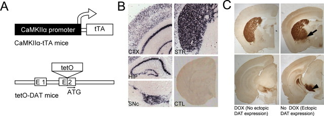
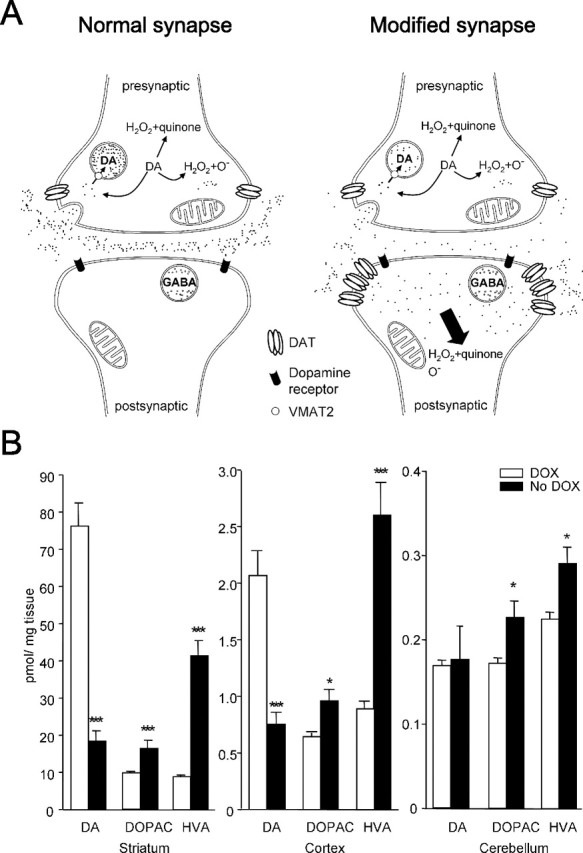
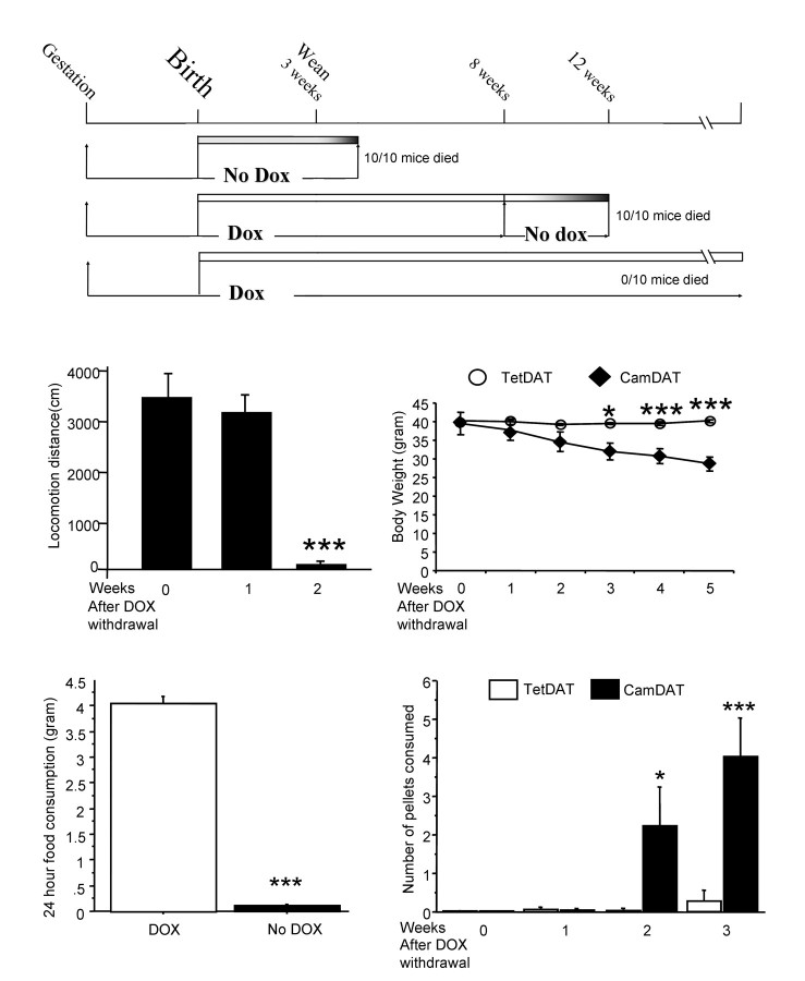
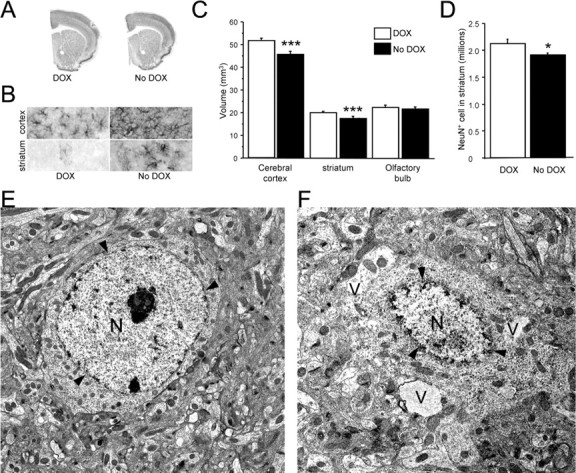
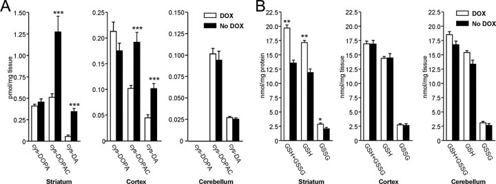
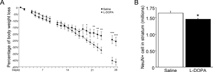
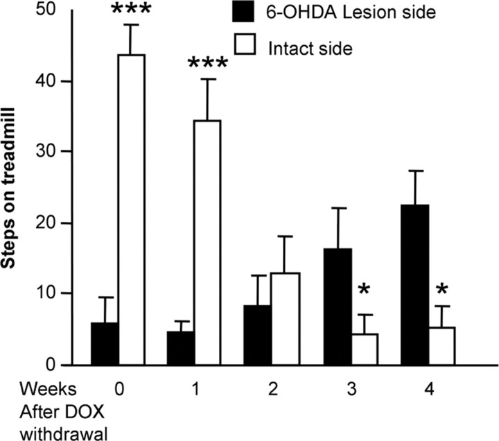
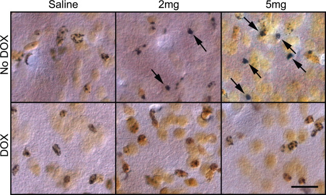
The role of dopamine as a vulnerability factor and a toxic agent in Parkinson's disease (PD) is still controversial, yet the presumed dopamine toxicity is partly responsible for the "DOPA-sparing" clinical practice that avoids using L-3,4-dihydroxyphenylalanine (L-DOPA), a dopamine precursor, in early PD. There is a lack of studies on animal models that directly isolate dopamine as one determining factor in causing neurodegeneration. To address this, we have generated a novel transgenic mouse model in which striatal neurons are engineered to take up extracellular dopamine without acquiring regulatory mechanisms found in dopamine neurons. These mice developed motor dysfunctions and progressive neurodegeneration in the striatum within weeks. The neurodegeneration was accompanied by oxidative stress, evidenced by substantial oxidative protein modifications and decrease in glutathione. Ultrastructural morphologies of degenerative cells suggest necrotic neurodegeneration. Moreover, L-DOPA accelerated neurodegeneration and worsened motor dysfunction. In contrast, reducing dopamine input to striatum by lesioning the medial forebrain bundle attenuated motor dysfunction. These data suggest that pathology in genetically modified striatal neurons depends on their dopamine supply. These neurons were also supersensitive to neurotoxin. A very low dose of 1-methyl-4-phenyl-1,2,3,6-tetrahydropyridine (5 mg/kg) caused profound neurodegeneration of striatal neurons, but not midbrain dopamine neurons. Our results provide the first in vivo evidence that chronic exposure to unregulated cytosolic dopamine alone is sufficient to cause neurodegeneration. The present study has significant clinical implications, because dopamine replacement therapy is the mainstay of PD treatment. In addition, our model provides an efficient in vivo approach to test therapeutic agents for PD.


Figures

Figure 1.
Figure 1.
Generation of transgenic mice with forebrain expression of DAT (CamDAT mice). A, Inducible forebrain DAT expression mice were generated by breeding forebrain-specific CaMKIIα-tTA mice and tetO-DAT mice. B, DAT mRNA expression was detected in the striatum (STR), cerebral cortex (CTX), hippocampus (HIP), olfactory bulb (not shown), and olfactory tubercle (not shown). Endogenous DAT mRNA was detected in SNc. In control mice (CTL), no DAT mRNA expression was detected in the forebrain. C, DAT proteins were detected in regions with mRNA expression as well as in globus pallidus (arrow) and substantia nigra pars reticulata (arrowhead) that are main striatum efferent nuclei. Top, Forebrain region shown in more rostral sections. Bottom, Midbrain region shown in more caudal sections.
Figure 2.
Figure 2.
Alternation of dopamine turnover in CamDAT mice. A, A schematic illustration of genetically engineered synapse in the striatum. Most dopamine released by presynaptic dopamine terminal is taken up by postsynaptic neurons expressing ectopic DAT. There is no VMAT2 expression in postsynaptic neurons. Uptaken dopamine is degraded in cytoplasm and produces quinine, H2O2, and other reactive species in postsynaptic neurons. B, After 3 weeks of DOX withdrawal, striatum and cortex dopamine content decreased and its metabolites, DOPAC and HVA, increased. In cerebellum, dopamine level was unchanged, whereas HVA and DOPAC levels increased. Note the differences in y-axis scales (n = 8 per group, unpaired t test, p < 0.001 for dopamine in striatum, p < 0.001 for DOPAC in striatum, p = 0.01 for HVA in striatum, p < 0.001 for dopamine in cortex, p = 0.018 for DOPAC in cortex, p < 0.001 for HVA in cortex, p = 0.015 for DOPAC in cerebellum, p = 0.013 for HVA in cerebellum).
Figure 3.
Figure 3.
Behavioral phenotypes of CamDAT mice. A, DOX treatment was required for the survival of CamDAT mice. B, In the open field, locomotor activity of CamDAT mice was normal 1 week after DOX withdrawal. However, their locomotor activity sharply declined in the second week of DOX withdrawal (n = 4 per group, repeated-measures ANOVA, p < 0.001). C, Progressive body weight loss in CamDAT mice after DOX withdrawal (n = 6, repeated-measures ANOVA, p < 0.001 for group × week interaction). D, Four weeks after DOX withdrawal, CamDAT mice in their home cages consumed very little regular rodent chow (n = 5 per group, individually housed, unpaired t test, p < 0.001). E, Two weeks after DOX withdrawal, when provided with easily accessible chocolate flavored pellets (20 mg each) on the cage floor, CamDAT mice ate more than TetDAT mice (n = 6 per group, repeated-measures ANOVA, p < 0.001 for group × week interaction).
Figure 4.
Figure 4.
Neuropathology in the forebrain of CamDAT mice. A, C, Four weeks after DOX withdrawal, the volumes of cerebral cortex and striatum decreased by 15 and 10% respectively, whereas volume of olfactory bulb was unchanged (n = 4 per group, unpaired t test, p < 0.001 for both striatum and cortex). B, Astrogliosis was detected by GFAP staining in the cerebral cortex and striatum. n = 6 per group. D, NeuN-positive cells in the striatum decreased by 10% (n = 4, unpaired t test, p = 0.04). E, Transmission electron microscopic image of a normal striatal neuron from a CamDAT mouse under DOX treatment. F, Abnormal morphologies were found in striatum of CamDAT mice after 4 weeks of DOX withdrawal. A severely affected neuron displayed vacuolization (V) and disintegration of the nucleus (N). Arrowheads indicate the nuclear envelope. Scale bar, 2 μm.
Figure 5.
Figure 5.
Oxidative stress in transgenic mice. A, Oxidation-induced protein modification by dopamine. Using HPLC, we found significant increases of cys-dopamine and cys-DOPAC in CamDAT mice after 3 weeks of DOX withdrawal. Similar increases were found in cerebral cortex. In contrast, no change was found in the cerebellum (n = 8 per group, unpaired t test, ***p < 0.001). B, Decreased glutathione content in brain tissues. Total glutathione (GSH+GSSG), GSH, and GSSG were 26% (p < 0.01), 29% (p < 0.01), and 27% (p < 0.05) lower, respectively, in striatum of CamDAT mice after 4 weeks of DOX withdrawal compared with control mice (n = 5 per group, unpaired t test, *p < 0.05, **p < 0.01). No change was found either in the cortex or cerebellum (p > 0.05, n = 5 per group).
Figure 6.
Figure 6.
l-DOPA accelerates neurodegeneration and body weight loss. Four weeks after DOX withdrawal, CamDAT mice were injected daily with 300 mg/kg l-DOPA plus 100 mg/kg benserazide, and body weight was monitored. A, l-DOPA-treated mice gradually lost more body weight than saline control (41.5% vs 25%, respectively, n = 5 per group, unpaired t test, p < 0.001). Day 0 indicates the first day of l-DOPA treatment. Blank data points indicate the days without treatment. B, l-DOPA accelerated neurodegeneration in striatum assayed by unbiased stereological cell counting (12% more loss of NeuN+ neurons in l-DOPA-treated mice, n = 4 per group, unpaired t test, p = 0.03). *p < 0.05, **p < 0.01, ***p < 0.001.
Figure 7.
Figure 7.
Motor dysfunction in CamDAT mice is dopamine supply dependent. DOX treatment was removed 1 month after unilateral 6-OHDA lesion, and stepping of the forelimb controlled by the intact side worsened progressively, dropping from 100 to 15% of baseline level during 4 weeks DOX withdrawal, whereas stepping of the forelimb controlled by the lesion side improved from 16 to 50% of baseline level (n = 5 per group, repeated-measures ANOVA, p < 0.001 for side × week interaction).
Figure 8.
Figure 8.
Low-dose MPTP causes neurodegeneration in forebrain. Using FD silver staining kit, degenerative neurons in striatum were found in an MPTP dose-dependent manner (2 mg/kg, 5 mg/kg). The degenerative neurons (arrows) are characterized by dense, dark silver precipitates in their cell bodies. This type of neuron is absent in saline and DOX groups. n = 3 per group. Scale bar, 20 μm.

References

    1. Agid Y, Ahlskog E, Albanese A, Calne D, Chase T, De Yebenes J, Factor S, Fahn S, Gershanik O, Goetz C, Koller W, Kurth M, Lang A, Lees A, Lewitt P, Marsden D, Melamed E, Michel PP, Mizuno Y, Obeso J, et al. Levodopa in the treatment of Parkinson's disease: a consensus meeting. Mov Disord. 1999;14:911–913. - PubMed
    1. Barzilai A, Daily D, Zilkha-Falb R, Ziv I, Offen D, Melamed E, Shirvan A. The molecular mechanisms of dopamine toxicity. Adv Neurol. 2003;91:73–82. - PubMed
    1. Bonifati V, Rizzu P, van Baren MJ, Schaap O, Breedveld GJ, Krieger E, Dekker MC, Squitieri F, Ibanez P, Joosse M, van Dongen JW, Vanacore N, van Swieten JC, Brice A, Meco G, van Duijn CM, Oostra BA, Heutink P. Mutations in the DJ-1 gene associated with autosomal recessive early-onset parkinsonism. Science. 2003;299:256–259. - PubMed
    1. Cappai R, Leck SL, Tew DJ, Williamson NA, Smith DP, Galatis D, Sharples RA, Curtain CC, Ali FE, Cherny RA, Culvenor JG, Bottomley SP, Masters CL, Barnham KJ, Hill AF. Dopamine promotes alpha-synuclein aggregation into SDS-resistant soluble oligomers via a distinct folding pathway. FASEB J. 2005;19:1377–1379. - PubMed
    1. Caudle WM, Richardson JR, Wang MZ, Taylor TN, Guillot TS, McCormack AL, Colebrooke RE, Di Monte DA, Emson PC, Miller GW. Reduced vesicular storage of dopamine causes progressive nigrostriatal neurodegeneration. J Neurosci. 2007;27:8138–8148. - PMC - PubMed

Publication types

MeSH terms

LinkOut - more resources