. 2008 Jan 9;28(2):483-90.
doi: 10.1523/JNEUROSCI.4067-07.2008.

Wild-type but not FAD mutant presenilin-1 prevents neuronal degeneration by promoting phosphatidylinositol 3-kinase neuroprotective signaling

Affiliations

Wild-type but not FAD mutant presenilin-1 prevents neuronal degeneration by promoting phosphatidylinositol 3-kinase neuroprotective signaling

Lia Baki et al. J Neurosci. .

Abstract

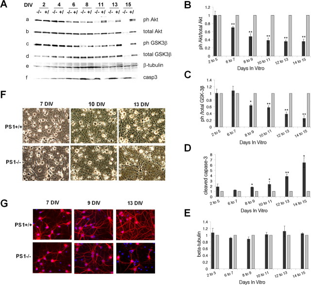
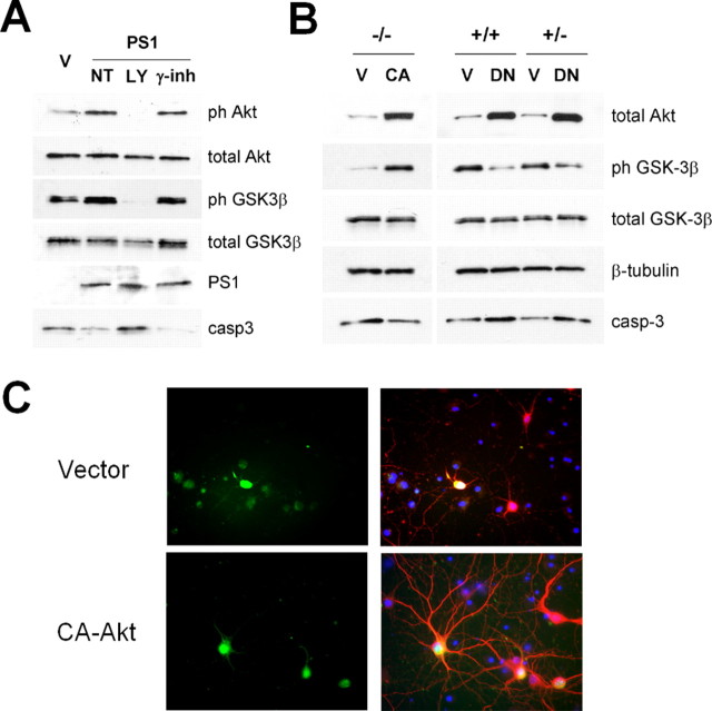
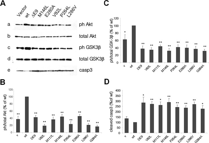
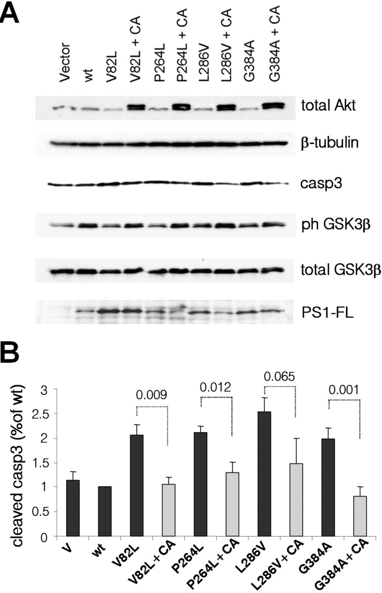
The role of presenilin-1 (PS1) in neuronal phosphatidylinositol 3-kinase (PI3K)/Akt signaling was investigated in primary neuronal cultures from wild-type (WT) and PS1 null (PS1-/-) embryonic mouse brains. Here we show that in PS1-/- cultures, the onset of neuronal maturation coincides with a decrease in the PI3K-dependent phosphorylation-activation of Akt and phosphorylation-inactivation of glycogen synthase kinase-3 (GSK-3). Mature PS1-/- neurons show increased activation of apoptotic caspase-3 and progressive degeneration preceded by dendritic retraction. Expression of exogenous WT PS1 or constitutively active Akt in PS1-/- neurons stimulates PI3K signaling and suppresses both caspase-3 activity and dendrite retraction. The survival effects of PS1 are sensitive to inhibitors of PI3K kinase but insensitive to gamma-secretase inhibitors. Familial Alzheimer disease (FAD) mutations suppress the ability of PS1 to promote PI3K/AKT signaling, prevent phosphorylation/inactivation of GSK-3 and promote activation of caspase-3. These mutation effects are reversed upon coexpression of constitutively active Akt. Together, our data indicate that the neuroprotective role of PS1 depends on its ability to activate the PI3K/Akt signaling pathway and that PS1 FAD mutations increase GSK-3 activity and promote neuronal apoptosis by inhibiting the function of PS1 in this pathway. These observations suggest that stimulation of PI3K/Akt signaling may be beneficial to FAD patients.


Figures

Figure 1.
Figure 1.
Impaired PI3K/Akt signaling and increased apoptosis in 13DIV PS1−/− neurons. A, Lysates from13DIV PS1+/+, PS1−/− and PS1+/− mouse primary neurons were analyzed for phosphorylated (Ser473) and total Akt (a, b) phosphorylated (Ser9) and total GSK-3β (c, d), cleaved (activated) caspase 3 (cap3) and PS1-CTF (33B10). B–D, Statistical analysis with each bar representing data from five to seven embryos. E, DAPI nuclear staining of PS1+/+ and PS1−/− neurons cultured on glass coverslips for 13 d. Fragmented nuclei are indicated by circles, nuclear fragments are indicated by arrows. F, Annexin V-CY3 staining of 13 DIV PS1+/+ and PS1−/− neurons. Asterisks are described in Materials and Methods.
Figure 2.
Figure 2.
Downregulation of PI3K/Akt signaling in PS1−/− neurons at the onset of neuronal maturation period. A, PS1−/− and PS1+/− mouse primary neurons were maintained in culture for the indicated periods. Lysates were analyzed as indicated. B–E, Statistical analysis with each bar representing data from at least three independent experiments (dark gray, PS1−/−; light gray, PS1+/−). Levels for ph-Akt and ph-GSK were normalized to the levels of total Akt and GSK respectively and data are presented as ratio of phosphorylated total protein. All values are expressed relative to the respective values of the PS1+/− samples. F, Phase contrast pictures of PS1+/+ and PS1−/− primary neurons cultured for 7, 10, and 13 d in vitro. G, MAP2 (red) immunostaining of PS1+/+ and PS1−/− mouse primary neurons cultured on coverslips for 7, 9, and 13 d. Nuclei (blue) were stained with DAPI.
Figure 3.
Figure 3.
Re-introduction of PS1 into PS1−/− neurons restores PI3K/Akt signaling and suppresses neuronal degeneration. A, PS1−/− primary neurons were infected with either HSV-vector virus (V), or HSV-PS1 (PS1) recombinant virus on their 10th day in culture. Lysates were analyzed as indicated. B, Statistical analysis of the results from four independent experiments. C, MAP2 (red) and DAPI (blue) staining of 13 DIV PS1−/− mouse primary neurons, infected with EGFP-expressing (green) HSV-EGFP-vector and HSV-EGFP-PS1 recombinant viruses.
Figure 4.
Figure 4.
PS1 rescues via PI3K/Akt signaling. A, PS1−/− primary neurons, infected with either HSV-vector virus (V), or HSV-PS1 (PS1) recombinant viruses were incubated in the absence (NT) or presence of LY294002 (LY) or the γ-secretase inhibitor l-685,458 (γ-inh). Lysates were analyzed as indicated. B, Lysates from PS1−/−, PS1+/+ and PS1+/− primary neurons, infected with HSV-EGFP-vector, HSV-EGFP-constitutively active Akt or HSV-EGFP dominant-negative Akt(DN) viruses were analyzed as indicated. C, MAP2 (red) and DAPI (blue) staining of 14 DIV PS1−/− mouse primary neurons, infected with EGFP-expressing (green) HSV-EGFP-vector and HSV-EGFP CA-Akt recombinant viruses.
Figure 5.
Figure 5.
PS1 FAD mutations impair neuronal PI3K/Akt signaling, leading to increased GSK-3 activity and apoptosis. A, PS1+/− primary neurons were infected with HSV-vector (V) or HSV recombinant viruses encoding WT PS1 (wt) or one of the PS1 FAD mutations shown on the top of the lanes. Lysates were analyzed as indicated. B–D, Statistical analysis with each mutation being analyzed in at least four (B, C) or at least three (D) independent experiments.
Figure 6.
Figure 6.
Constitutively active Akt reverses the effects of PS1 FAD mutations. A, PS1+/− primary neurons were infected with HSV-vector (V) or HSV recombinant viruses encoding WT PS1 (wt) or one of the PS1 FAD mutations shown. CA refers to coinfection with HSV-mutant-PS1 viruses plus HSV-CA-Akt viruses (+CA). Lysates were analyzed as indicated. B, Statistical analysis of the data from three independent experiments. The numbers indicate statistical significance.

References

    1. Baki L, Shioi J, Wen P, Shao Z, Schwarzman A, Gama-Sosa M, Neve R, Robakis NK. PS1 activates PI3K thus inhibiting GSK-3 activity and tau overphosphorylation: effects of FAD mutations. EMBO J. 2004;23:2586–2596. - PMC - PubMed
    1. Bhat RV, Budd Haeberlein SL, Avila J. Glycogen synthase kinase 3: a drug target for CNS therapies. J Neurochem. 2004;89:1313–1317. - PubMed
    1. Brunet A, Datta SR, Greenberg ME. Transcription-dependent and -independent control of neuronal survival by the PI3K-Akt signaling pathway. Curr Opin Neurobiol. 2001;11:297–305. - PubMed
    1. Chen G, Bower KA, Ma C, Fang S, Thiele CJ, Luo J. Glycogen synthase kinase 3β (GSK3beta) mediates 6-hydroxydopamine-induced neuronal death. FASEB J. 2004;18:1162–1164. - PubMed
    1. Chen Y, Bodles AM, McPhie DL, Neve RL, Mrak RE, Griffin WS. APP-BP1 inhibits Abeta42 levels by interacting with Presenilin-1. Mol Neurodegener. 2007;2:3. - PMC - PubMed

Publication types

MeSH terms