

Case Reports
doi: 10.3174/ajnr.A0924.
Epub 2008 Jan 9.
Malignant transformation of a lipomyelocele into a rhabdomyosarcoma?
Affiliations
- PMID: 18184836
- PMCID: PMC8118878
- DOI: 10.3174/ajnr.A0924
Item in Clipboard
Case Reports
Malignant transformation of a lipomyelocele into a rhabdomyosarcoma?
AJNR Am J Neuroradiol.
2008 Mar.
Abstract
We report the unusual transformation of a lipomyelocele to a rhabdomyosarcoma in a 3-year-old boy. A lipomyelocele is not considered pre-malignant, but the possibility of developing such a tumor may represent another reason for close neurological follow-up in patients with spinal dysraphism.
Figures

Photograph of a complex ulcerating hemangioma over the sacrum in a full-term neonate on day 1.
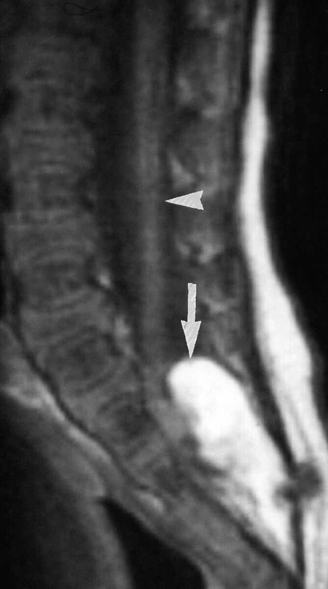
Sagittal T1-weighted MR image obtained at 3 months confirms the sonographic finding of the tethered spinal cord (arrowhead) ending in a lipomyelocele (arrow).

A, Sagittal T1-weighted MR image at 3 years shows a new large homogeneous mass of low signal intensity (arrow), apparently arising from the lipomyelocele and distal spinal cord within the spinal canal. B, Postcontrast T1-weighted sagittal MR image shows multiple foci (arrowheads) of high signal intensity in the distal thoracic and lumbar spinal cord consistent with metastatic seeding. C, Sagittal postcontrast T1-weighted MR image demonstrates multiple foci (arrowheads) of high signal intensity in the brain stem and cervical and upper thoracic spinal cord, consistent with metastatic seeding.

Resection of this sacral mass revealed a pleomorphic malignant spindle cell tumor at pathology, consistent with a rhabdomyosarcoma (hematoxylin-eosin).
References
-
- Unsinn KM, Geley T, Freund MC, et al. US of the spinal cord in newborns: spectrum of normal findings, variants, congenital anomalies, and acquired diseases. Radiographics 2000;20:923–38 - PubMed
-
- Rossi A. Imaging in spine and spinal cord malformations. Eur J Radiol 2004;50:177–200 - PubMed
-
- Freyer DR, Hutchinson RJ, McKeever PE. Primary primitive neuroectodermal tumor of the spinal cord associated with neural tube defect. Pediatr Neurosci 1989;15:181–87 - PubMed
-
- Thorp RH. Carcinoma associated with myelomeningocele: case report. J Neurosurg 1967;27:446–48 - PubMed
Publication types
MeSH terms
LinkOut - more resources
Full Text Sources