

Perfusion imaging of brain tumors using arterial spin-labeling: correlation with histopathologic vascular density
- PMID: 18184842
- PMCID: PMC7978189
- DOI: 10.3174/ajnr.A0903
Perfusion imaging of brain tumors using arterial spin-labeling: correlation with histopathologic vascular density
Abstract
Background and purpose: We investigated the relationship between tumor blood-flow measurement based on perfusion imaging by arterial spin-labeling (ASL-PI) and histopathologic findings in brain tumors.
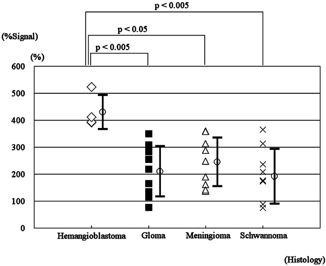
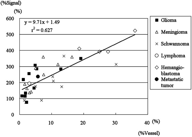
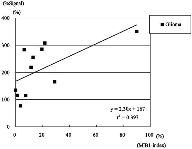
Materials and methods: We used ASL-PI to examine 35 patients with brain tumors, including 11 gliomas, 9 meningiomas, 9 schwannomas, 1 diffuse large B-cell lymphoma, 4 hemangioblastomas, and 1 metastatic brain tumor. As an index of tumor perfusion, the relative signal intensity (SI) of each tumor (%Signal intensity) was determined as a percentage of the maximal SI within the tumor per averaged SI within normal cerebral gray matter on ASL-PI. Relative vascular attenuation (%Vessel) was determined as the total microvessel area per the entire tissue area on CD-34-immunostained histopathologic specimens. MIB1 indices of gliomas were also calculated. The differences in %Signal intensity among different histopathologic types and between high- and low-grade gliomas were compared. In addition, the correlations between %Signal intensity and %Vessel or MIB1 index were evaluated in gliomas.
Results: Statistically significant differences in %Signal intensity were observed between hemangioblastomas versus gliomas (P < .005), meningiomas (P < .05), and schwannomas (P < .005). Among gliomas, %Signal intensity was significantly higher for high-grade than for low-grade tumors (P < .05). Correlation analyses revealed significant positive correlations between %Signal intensity and %Vessel in 35 patients, including all 6 histopathologic types (rs = 0.782, P < .00005) and in gliomas (rs = 0.773, P < .05). In addition, in gliomas, %Signal intensity and MIB1 index were significantly positively correlated (rs = 0.700, P < .05).
Conclusion: ASL-PI may predict histopathologic vascular densities of brain tumors and may be useful in distinguishing between high- and low-grade gliomas and in differentiating hemangioblastomas from other brain tumors.
Figures


References
-
- Chalela JA, Alsop DC, Gonzalez-Atavales JB, et al. Magnetic resonance perfusion imaging in acute ischemic stroke using continuous arterial spin labeling. Stroke 2000;31:680–87 - PubMed
-
- Kimura H, Kado H, Koshimoto Y, et al. Multislice continuous arterial spin-labeled perfusion MRI in patients with chronic occlusive cerebrovascular disease: a correlative study with CO2 PET validation. J Magn Reson Imaging 2005;22:189–98 - PubMed
-
- Detre JA, Alsop DC, Vives LR, et al. Noninvasive MRI evaluation of cerebral blood flow in cerebrovascular disease. Neurology 1998;50:633–41 - PubMed
-
- Alsop DC, Detre JA, Grossman M. Assessment of cerebral blood flow in Alzheimer's disease by spin-labeled magnetic resonance imaging. Ann Neurol 2000;47:93–100 - PubMed
Publication types
MeSH terms
Substances
LinkOut - more resources
Full Text Sources
Other Literature Sources
Medical
Research Materials
Miscellaneous