. 2008 Feb;19(2):321-8.
doi: 10.1681/ASN.2007040481. Epub 2008 Jan 9.

Asialoerythropoietin prevents contrast-induced nephropathy

Affiliations

Asialoerythropoietin prevents contrast-induced nephropathy

Yukiyo Yokomaku et al. J Am Soc Nephrol. 2008 Feb.

Abstract

Strategies to prevent contrast-induced nephropathy (CIN) are suboptimal. Erythropoietin was recently found to be cytoprotective in a variety of nonhematopoietic cells, so it was hypothesized that the nonhematopoietic erythropoietin derivative asialoerythropoietin would prevent CIN. Nephropathy was induced in rats by injection of the radiocontrast medium Ioversol in addition to inhibition of prostaglandin and nitric oxide synthesis. Administration of a single dose of asialoerythropoietin before the induction of nephropathy significantly attenuated the resulting renal dysfunction and histologic renal tubular injury. Contrast-induced apoptosis of renal tubular cells was inhibited by asialoerythropoietin both in vivo and in vitro, and this effect was blocked by a Janus kinase 2 (JAK2) inhibitor in vitro. Furthermore, phospho-JAK2/signal transducer and activator of transcription 5 (STAT5) and heat-shock protein 70 increased after injection of asialoerythropoietin, suggesting that the effects of asialoerythropoietin may be mediated by the activation of the JAK2/STAT5 pathway. Overall, these findings suggest that asialoerythropoietin may have potential as a new therapeutic approach to prevent CIN given its ability to preserve renal function and directly protect renal tissue.


Figures

Figure 1.
Figure 1.
EPO or asialoEPO attenuates the contrast medium–induced morphologic changes. The marked tubular injuries caused by Ioversol (C and H) are diminished by pretreatment with EPO (D and I) or asialoEPO (E and J). Representative photomicrographs of HE-stained kidney sections are presented. (A and F) Control rat. (B and G) Control rat pretreated with asialoEPO. (C and H) Rat treated with Ioversol. (D and I) Rat treated with Ioversol after EPO treatment. (E and J) Rat treated with Ioversol after asialoEPO treatment. Figures are representative of four to 10 rats in each group. Magnifications: ×100 in A through E; ×400 in F through J.
Figure 2.
Figure 2.
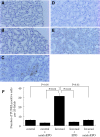
Administration of EPO or asialoEPO inhibits renal tubular apoptosis caused by contrast medium. The induction of Ioversol-induced nephropathy increases the number of TUNEL-positive renal tubular cells, but the number is markedly decreased by preadministration of EPO or asialoEPO. Representative photomicrographs of TUNEL-stained kidney sections from control rat (A), control rat pretreated with asialoEPO (B), rat treated with Ioversol (C), rat treated with Ioversol after EPO treatment (D), and rat treated with Ioversol after asialoEPO treatment (E). Figures are representative of at least four rats in each group. Quantitative analysis of the number of TUNEL-positive cells per 20 random fields also reveals that administration EPO or asialoEPO significantly inhibits renal tubular apoptosis caused by Ioversol (F). Data are means ± SEM for four to 10 rats in each group. Magnification, ×400.
Figure 3.
Figure 3.
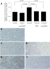
Administration of EPO or asialoEPO inhibits the activation of caspase-3 caused by contrast medium. In homogenates of rat kidneys 24 h after injection of Ioversol, caspase-3 activity was significantly increased compared with control rats. (A) EPO or asialoEPO administration significantly reduced caspase-3 activities. Data are means ± SE for four to 10 rats in each group. The exposure to Ioversol also increases cytoplasmic staining in the renal tubular cells at 24 h after exposure, but the staining for caspase-3 is markedly reduced in the Ioversol-treated groups that were pretreated with EPO or asialoEPO (B through F). Representative photomicrographs of kidney sections immunohistochemically stained with anti–active cleaved caspase-3 antibody from control rat (B), control rat pretreated with asialoEPO (C), rat treated with Ioversol (D), rat treated with Ioversol after EPO treatment (E), and rat treated with Ioversol after asialoEPO treatment (F). Magnification, ×100.
Figure 4.
Figure 4.
EPO or asialoEPO directly inhibits Ioversol-induced apoptosis in cultured LLC-PK1 cells. The cells were incubated with Ioversol for 30 min with or without pretreatment with EPO or asialoEPO for 1 h, and the activities of caspase-3 were measured at 24 h after Ioversol exposure. Data are means ± SEM (n = 4).
Figure 5.
Figure 5.
Administration of asialoEPO or EPO activates intracellular signals in cultured proximal tubular cells. Cultured LLC-PK1 cells were incubated with asialoEPO (25 ng/ml) or EPO (25 ng/ml). Phosphorylated/total JAK2, STAT5, and HSP70 expressions in the LLC-PK1 cells were determined by immunoblot analysis. (A) Phosphorylated and total JAK2 and STAT5 after 5-min exposure to asialoEPO or EPO were determined with immunoblot analysis with the indicated antibody. The expressions of HSP70 and β-actin after 1-h exposure were also determined with immunoblot analysis. Blots are representative of three independent experiments. (B) Each set of immunoblots was quantitatively evaluated with densitometric analysis. (C) Pretreatment with JAK2 inhibitor (AG490; 50 μM) for 1 h significantly reversed the inhibitory effects of asialoEPO on Ioversol-induced caspase-3 activation.
Figure 6.
Figure 6.
Administration of asialoEPO or EPO activates intracellular signals in rat kidneys in vivo. After bolus injection of asialoEPO (80 ng/g) or EPO (80 ng/g) in rats, the renal homogenates were subjected to immunoblot analysis. (A) Phosphorylated/total JAK2 and STAT5 after 5-min exposure to asialoEPO or EPO and the expression of HSP70 after 1-h exposure to asialoEPO or EPO in the kidney samples. Blots are representative of at least four rats. (B) Each set of immunoblots was quantitatively evaluated with densitometric analysis.

References

    1. Finn WF: The clinical and renal consequences of contrast-induced nephropathy. Nephrol Dial Transplant 21: i2–i10, 2006 - PubMed
    1. Tepel M, Aspelin P, Lameire N: Contrast-induced nephropathy: A clinical and evidence-based approach. Circulation 113: 1799–1806, 2006 - PubMed
    1. Mehran R, Nikolsky E: Contrast-induced nephropathy: Definition, epidemiology, and patients at risk. Kidney Int Suppl 11–15, 2006 - PubMed
    1. Goldenberg I, Matetzky S: Nephropathy induced by contrast media: Pathogenesis, risk factors and preventive strategies. CMAJ 172: 1461–1471, 2005 - PMC - PubMed
    1. Briguori C, Marenzi G: Contrast-induced nephropathy: Pharmacological prophylaxis. Kidney Int Suppl 30–38, 2006 - PubMed

MeSH terms