. 2007 Sep;4(3):507-528.
doi: 10.1142/S0219843607001138.

A PHYSIOLOGIST'S PERSPECTIVE ON ROBOTIC EXOSKELETONS FOR HUMAN LOCOMOTION

Affiliations

A PHYSIOLOGIST'S PERSPECTIVE ON ROBOTIC EXOSKELETONS FOR HUMAN LOCOMOTION

Daniel P Ferris et al. Int J HR. 2007 Sep.

Abstract

Technological advances in robotic hardware and software have enabled powered exoskeletons to move from science fiction to the real world. The objective of this article is to emphasize two main points for future research. First, the design of future devices could be improved by exploiting biomechanical principles of animal locomotion. Two goals in exoskeleton research could particularly benefit from additional physiological perspective: 1) reduction in the metabolic energy expenditure of the user while wearing the device, and 2) minimization of the power requirements for actuating the exoskeleton. Second, a reciprocal potential exists for robotic exoskeletons to advance our understanding of human locomotor physiology. Experimental data from humans walking and running with robotic exoskeletons could provide important insight into the metabolic cost of locomotion that is impossible to gain with other methods. Given the mutual benefits of collaboration, it is imperative that engineers and physiologists work together in future studies on robotic exoskeletons for human locomotion.


Figures
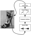
Figure 1
Figure 1
A pneumatically powered ankle-foot orthosis using proportional myoelectric control. Surface electrodes record the electromyography signal from the muscle of interest (soleus in this case) and send it to a computer for processing. The computer applies filters, a threshold, and a gain to generate a proportional control signal regulating air pressure in the artificial pneumatic muscle. Details are available in previous publications , , .
Figure 2
Figure 2
Ten subjects practiced walking with a single powered ankle-foot orthosis under soleus proportional myoelectric control. Subjects walked on a treadmill at 1.25 m/s for 55 minutes: 10 minutes with the orthosis unpowered (baseline), 30 minutes with the orthosis powered (powered), and 15 minutes with the orthosis unpowered again (post). Subjects completed two training sessions, three days apart (Day 1 and Day 2). A) Soleus root mean square electromyography (RMS EMG) during stance was normalized for each subject, averaged for each minute, and the mean value for each minute was calculated across all subjects (mean ± standard deviation, black circles and grey shading). Horizontal bars indicate steady state ranges. B) Ankle kinematic profiles and soleus electromyography profiles are displayed across training. Average data are shown for ankle joint kinematic profiles, soleus electromyography profiles. Within thirty minutes on Day 1, subjects returned to normal gait kinematics by reducing soleus muscle activation. On Day 2, subjects demonstrated a clear motor memory of orthosis dynamics. Curves are means across all subjects and the vertical bars indicate timing of the stance-swing transition. Data are from Gordon and Ferris
Figure 3
Figure 3
A comparison of locomotor adaptation to unilateral powered ankle-foot orthoses using two different controllers. Soleus electromyography root mean square (EMG RMS) activity is shown for each minute as mean ± 2 standard deviations across all subjects for each controller. Soleus proportional myoelectric control is shown in grey, and foot switch control is shown in black. Horizontal bars indicate steady state values for each controller (dark grey for footswitch, light grey for myoelectric control). When the orthosis is turned on by placement of the forefoot on the ground (footswitch control), subjects exhibit a smaller decrease in soleus muscle recruitment compared to proportional myoelectric control. Data are from Cain et al .
Figure 4
Figure 4
Orthosis mechanical power walking under soleus proportional myoelectric control. Grey curves are the mean ± standard deviation for all subjects during the first minute of testing on Day 1. Black curves are the mean ± standard deviation for all subjects during minute 30 on Day 2. By the end of Day 2, the orthosis produced almost exclusively positive mechanical power, which was focused at the end of stance. The vertical black line represents the stance-swing transition timing in the gait cycle. Data are from Gordon and Ferris .

Similar articles

Cited by

References

    1. Hughes J. Powered lower limb orthotics in paraplegia. Paraplegia. 1972;9:191–193. - PubMed
    1. Vukobratovic M, Hristic D, Stojiljkovic Z. Development of active anthropomorphic exoskeletons. Med Biol Eng. 1974;12:66–80. - PubMed
    1. Seireg A, Grundman JG. Design of a multitask exoskeletal walking device for paraplegics. In: Ghista DN, editor. Biomechanics of Medical Devices. Marcel Dekker, Inc; New York: 1981. pp. 569–639.
    1. Townsend MA, Lepofsky RJ. Powered walking machine prosthesis for paraplegics. Med Biol Eng. 1976;14:436–444. - PubMed
    1. Vukobratovic M. Humanoid robotics - past, present state, future. Int J Human Robot. 2007 in press.

LinkOut - more resources