

Clear cell colitis: a form of microscopic colitis in children
- PMID: 18186560
- PMCID: PMC2675119
- DOI: 10.3748/wjg.14.231
Clear cell colitis: a form of microscopic colitis in children
Abstract
Aim: To describe a new clinical and pathological subtype of microscopic colitis in children.
Methods: A selected group of children with abdominal pain, constipation and/or diarrhoea showing discrete or no macroscopic abnormalities on endoscopy was described.
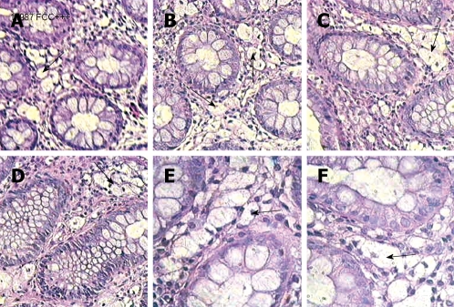
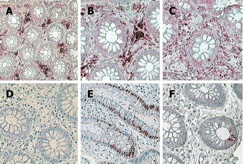
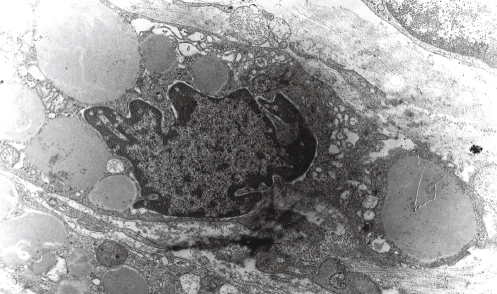
Results: Multiple biopsies of colon showed large mononuclear clear cells in lamina propria of mucous membrane provided that good quality histological sections were performed and observed under a higher magnification. Otherwise, they could be misinterpreted as artefacts. Their presence in routine histology might suggest a systemic storage disease (Whipple's disease), and neuronal intestine dysplasia. Using immunohistochemical staining and electron microscopy we confirmed their origin from CD68 positive mononuclear macrophages.
Conclusion: The presence of large clear cells is a constant microscopic feature. Failure of transient large bowel stationary macrophages plays a role in the pathogenesis of this benign microscopic clear cell colitis, sometimes coexisting with allergy.
Figures
References
-
- Mashako MN, Sonsino E, Navarro J, Mougenot JF, Gargouri A, Boige N, Cezard JP. Microscopic colitis: a new cause of chronic diarrhea in children? J Pediatr Gastroenterol Nutr. 1990;10:21–26. - PubMed
-
- Domizio P. Pathology of chronic inflammatory bowel disease in children. Baillieres Clin Gastroenterol. 1994;8:35–63. - PubMed
-
- da Silva JG, De Brito T, Cintra Damiao AO, Laudanna AA, Sipahi AM. Histologic study of colonic mucosa in patients with chronic diarrhea and normal colonoscopic findings. J Clin Gastroenterol. 2006;40:44–48. - PubMed
-
- Pardi DS, Smyrk TC, Tremaine WJ, Sandborn WJ. Microscopic colitis: a review. Am J Gastroenterol. 2002;97:794–802. - PubMed
MeSH terms
LinkOut - more resources
Full Text Sources
