. 2008 Jan 10:9:1.
doi: 10.1186/1471-2121-9-1.

The role of Drosophila Merlin in spermatogenesis

Affiliations

The role of Drosophila Merlin in spermatogenesis

Natalia V Dorogova et al. BMC Cell Biol. .

Abstract

Background: Drosophila Merlin, the homolog of the human Neurofibromatosis 2 (NF2) gene, is important for the regulation of cell proliferation and receptor endocytosis. Male flies carrying a Mer3 allele, a missense mutation (Met177-->Ile) in the Merlin gene, are viable but sterile; however, the cause of sterility is unknown.

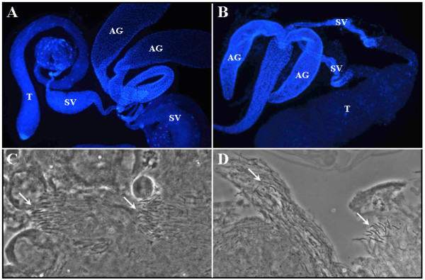
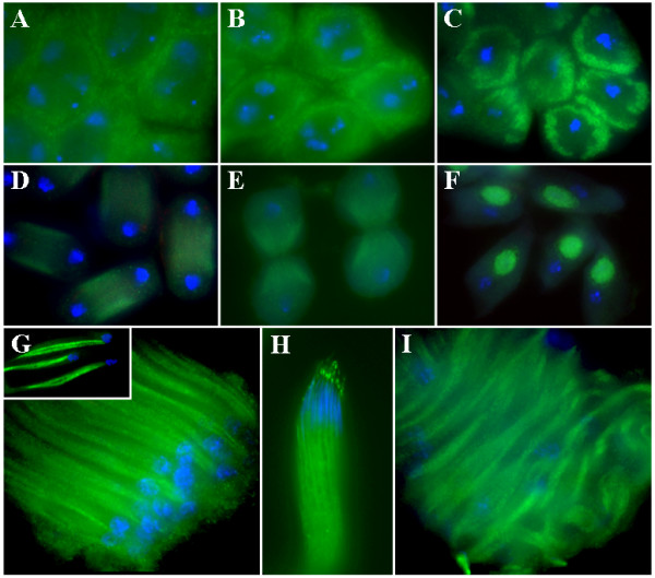
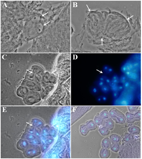
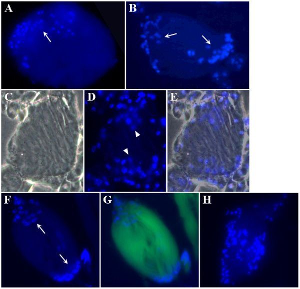
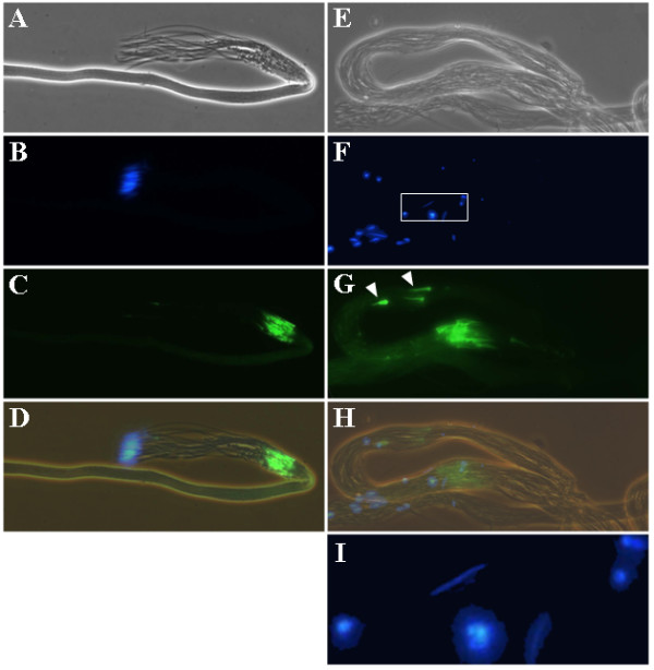
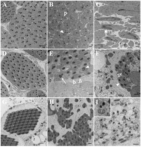
Results: Testis examination reveals that hemizygous Mer3 mutant males have small seminal vesicles that contain only a few immotile sperm. By cytological and electron microscopy analyses of the Mer3, Mer4 (Gln170-->stop), and control testes at various stages of spermatogenesis, we show that Merlin mutations affect meiotic cytokinesis of spermatocytes, cyst polarization and nuclear shaping during spermatid elongation, and spermatid individualization. We also demonstrate that the lethality and sterility phenotype of the Mer4 mutant is rescued by the introduction of a wild-type Merlin gene. Immunostaining demonstrates that the Merlin protein is redistributed to the area associated with the microtubules of the central spindle in telophase and its staining is less in the region of the contractile ring during meiotic cytokinesis. At the onion stage, Merlin is concentrated in the Nebenkern of spermatids, and this mitochondrial localization is maintained throughout sperm formation. Also, Merlin exhibits punctate staining in the acrosomal region of mature sperm.

Conclusion: Merlin mutations affect spermatogenesis at multiple stages. The Merlin protein is dynamically redistributed during meiosis of spermatocytes and is concentrated in the Nebenkern of spermatids. Our results demonstrated for the first time the mitochondrial localization of Merlin and suggest that Merlin may play a role in mitochondria formation and function during spermatogenesis.


Figures

Figure 1
Figure 1
Morphological examination of the testis tissues from young wild-type (panels A and C) and Mer3 (panels B and D) males. The control FM6 and Mer3 mutant males were obtained according to Materials and Methods. The DAPI-stained testis from the control male contained a bulging seminal vesicle (SV) (A). In contrast, a shriveled seminal vesicle was seen in the Mer3 testis (B). T, testis; AG, accessory gland. Acetic acid/orcein staining revealed that the testis tissue from the control male contained well-organized sperm bundles (arrows) (C), while the Mer3 testis had fewer sperm in a more disorganized bundle (arrows) (D).
Figure 2
Figure 2
Intracellular distribution of the Merlin protein at various stages of spermatogenesis. In the control cyst, Merlin was detected in the cellular cortex of spermatocytes (A). In prometaphase (B) and metaphase (C) of meiosis, the cortical localization of Merlin became more evident. In telophase (D), Merlin redistributed and accumulated in the area associated with the microtubules of the central spindle. During cytokinesis (E), Merlin staining remained associated with the microtubules but was less intense in the region of the contractile ring. In the onion-stage spermatids (F), Merlin was highly concentrated in the Nebenkern. This localization pattern was maintained through the comet stage of spermatid elongation (G). The insert in panel G shows intense Merlin staining in the two subunits of Nebenkern in spermatids. In the control cyst, containing mature sperm, bright Merlin staining was also seen as a punctate dot in the acrosomal region (H). Merlin staining was still detected in the Mer3 cyst at the comet stage; however, sperm nuclei were scattered throughout the cyst, and the arrangement of spermatids was irregular (I).
Figure 3
Figure 3
Abnormalities observed during meiosis of Mer3 spermatocytes: a spermatid containing two nuclei of equal size (arrows) and two Nebenkerns (A), tripolar spindle in a spermatocyte going through the second meiotic division (B), and four-polar spindle in a spermatocyte undergoing the second meiotic division (C-E). Panel C shows the phase contrast image, panel D the DAPI-stained nuclei, and panel E the merged image. Arrow points to the central spindle midzone [29]. For comparison, wild-type meiotic cells in telophase were shown (F).
Figure 4
Figure 4
Difference in nuclear grouping during cyst polarization between the control and Merlin mutant spermatids. Dissected testes from the control FM6 (A), Mer3 (B-E), and Mer4 (F-H) males were stained with DAPI and examined as described before. Note that while the spermatid nuclei were grouped in one area (arrow) of the control cyst (A), the spermatid nuclei in the Mer3 (B-E) and Mer4 (F-G) cysts were seen as two diffuse groups (arrows). In some cases, the spermatid nuclei were scattered in the mutant cyst (H). Panel C shows a phase-contrast image of a Mer3 cyst, panel D displays the same cyst stained with DAPI, and panel E represents a merged image. The cyst shown in panels F and G was obtained from a male carrying the Mer4 mutation and a GFP marker as described in Materials and Methods.
Figure 5
Figure 5
Spermatid individualization in the control (A-D) and Mer3 (E-I) cysts. Panels A and E illustrate the phase-contrast view of a control or Mer3 cyst, respectively. Panels B and F show the location of the DAPI-stained sperm nuclei. Panels C and G display the sites (arrowheads) and orientation of the actin cones as visualized by FITC-conjugated phalloidin staining. Panels D and H represent merged images. Panel I is an enlarged view of the rectangular area denoted in panel F.
Figure 6
Figure 6
Ultrastructural analysis of the control and Merlin mutant cysts during the elongation and individualization stages. (A-C) Sections of the cysts from the control FM6 (A), Mer3 (B), or Mer4 (C) testis at the elongation stage. (A) A dark paracrystalline body was seen within the major mitochondrial derivative in the control spermatid. Bar = 2 μm. (B) Some of the spermatids in the Mer3 cyst contained two paracrystalline bodies (filled arrowhead points to one example). Also, some spermatids had two axonemes (open arrowhead). Bar = 0.5 μm. (C) Two paracrystalline bodies within the major mitochondrial derivative were frequently seen in the elongating spermatids of the Mer4 cyst. Bar = 0.5 μm. (D-F) Cysts from the control FM6 (D), Mer3 (E), or Mer4 (F) testis at the individualization stage. (D) The association of axoneme with the mitochondrial derivatives was seen in the spermatids of the control cyst. Bar = 2 μm. (E) Spermatids in the Mer3 cyst might contain two abnormally-shaped (filled arrowhead) or three (arrow) paracrystalline bodies together with one axoneme, or have two paracrystalline-filled Nebenkerns but without the axoneme (open arrowhead). Bar = 0.5 μm. (F) Spermatids with multiple Nebenkerns with (open arrowhead) or without (filled arrowhead) axonemes together with cytoplasmic fragmentation were seen in the Mer4 cyst. Bar = 0.5 μm. (G-I) Cysts from the control FM6 (G), Mer3 (H), or Mer4 (I) testis at the late stage of individualization. (G) Individualized spermatids in the control cyst displayed a highly-ordered axoneme-Nebenkern pair. Bar = 0.5 μm. (H) The spermatids in the Mer3 cyst were poorly arranged and some of them appeared to be fused together. Bar = 0.5 μm. (I) The Mer4 cyst showed a complete destruction of spermatid individualization, resulting in empty spermatids with or without axoneme or Nebenkern. Insert illustrates the structure of axoneme appeared to be intact. Bar = 1 μm. Bar in the insert = 0.2 μm.

References

    1. Lindsley DL, Tokuyasu KT. Spermatogenesis. In: Ashburner M, Wright TR, editor. Genetics and Biology of Drosophila. 2. Academic Press, NY, NY; 1980. pp. 225–294.
    1. Fuller M. Spermatogenesis. In: Bate M, Arias AM, editor. The Development of Drosophila melanogaster. Cold Spring Harbor Laboratory Press, NY; 1993. pp. 71–147.
    1. Cross DP, Shellenbargerr DL. The dynamics of Drosophila melanogaster spermatogenesis in in vitro cultures. J Embryol Exp Morph. 1979;53:345–351. - PubMed
    1. Fabrizio J, Hime G, Lemmon S, Bazinet C. Genetic dissection of sperm individualization in Drosophila melanogaster. Development. 1998;125:1833–1843. - PubMed
    1. Fuller MT. Genetic control of cell proliferation and differentiation in Drosophila spermatogenesis. Semin Cell Dev Biol. 1998;9:433–444. doi: 10.1006/scdb.1998.0227. - DOI - PubMed

Publication types