Identification of platelet-derived growth factor D in human chronic allograft nephropathy
- PMID: 18187181
- PMCID: PMC2703673
- DOI: 10.1016/j.humpath.2007.07.008
Identification of platelet-derived growth factor D in human chronic allograft nephropathy
Abstract
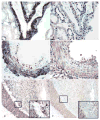
Chronic allograft nephropathy (CAN), a descriptive term denoting chronic scarring injury of the renal parenchyma and vasculature in allograft kidneys arising from various etiologies including chronic rejection, is the most common cause of late allograft failure, but mediators of this progressive injury largely remain unknown. We hypothesized that platelet-derived growth factor D (PDGF-D) and its specific receptor PDGF-Rbeta may be an important mediator in the pathogenesis of CAN and, hence, sought to identify its expression in this setting. Allograft nephrectomies demonstrating CAN, obtained from patients with irreversible transplant kidney failure (n = 15), were compared with renal tissues without prominent histopathological abnormalities (n = 18) and a series of renal allograft biopsies demonstrating acute vascular rejection (AVR) (n = 12). Antibodies to PDGF-D and PDGF-Rbeta were used for immunohistochemistry. Double and triple immunohistochemistry was used to identify cell types expressing PDGF-D. PDGF-D was widely expressed in most neointimas in arteries exhibiting the chronic arteriopathy of CAN and only weakly expressed in a small proportion of sclerotic arteries in the other 2 groups. Double and triple immunolabeling demonstrated that the neointimal cells expressing PDGF-D were alpha-smooth muscle actin-expressing cells, but not infiltrating macrophages or endothelial cells. PDGF-Rbeta expression evaluated in serial sections was localized to the same sites where neointimal PDGF-D was expressed. PDGF-Rbeta was expressed in interstitial cells more abundantly in the CAN group compared with the normal and AVR groups, without demonstrable colocalization of PDGF-D. PDGF-D is present in the neointima of the arteriopathy of CAN, where it can engage PDGF-Rbeta to promote mesenchymal cell migration, proliferation, and neointima formation. PDGF-D may engage the PDGF-Rbeta to promote interstitial injury in chronic allograft injury, but its sources within the interstitium were unidentified.
Conflict of interest statement
Figures


Similar articles
-
Identification of platelet-derived growth factor A and B chains in human renal vascular rejection.Am J Pathol. 1996 Feb;148(2):439-51. Am J Pathol. 1996. PMID: 8579107 Free PMC article.
-
Expression of PDGF alpha-receptor in renal arteriosclerosis and rejecting renal transplants.J Am Soc Nephrol. 1998 Feb;9(2):211-23. doi: 10.1681/ASN.V92211. J Am Soc Nephrol. 1998. PMID: 9527397
-
The effect of acute rejection and cyclosporin A-treatment on induction of platelet-derived growth factor and its receptors during the development of chronic rat renal allograft rejection.Transplantation. 2002 Feb 27;73(4):506-11. doi: 10.1097/00007890-200202270-00003. Transplantation. 2002. PMID: 11889420
-
Early induction of platelet-derived growth factor ligands and receptors in acute rat renal allograft rejection.Transplantation. 2001 Jul 15;72(1):31-7. doi: 10.1097/00007890-200107150-00009. Transplantation. 2001. PMID: 11468531
-
A new look at platelet-derived growth factor in renal disease.J Am Soc Nephrol. 2008 Jan;19(1):12-23. doi: 10.1681/ASN.2007050532. Epub 2007 Dec 12. J Am Soc Nephrol. 2008. PMID: 18077793 Review.
Cited by
-
Targeting CTGF, EGF and PDGF pathways to prevent progression of kidney disease.Nat Rev Nephrol. 2014 Dec;10(12):700-11. doi: 10.1038/nrneph.2014.184. Epub 2014 Oct 14. Nat Rev Nephrol. 2014. PMID: 25311535 Review.
-
VEGF/VEGFR2 and PDGF-B/PDGFR-β expression in non-metastatic renal cell carcinoma: a retrospective study in 1,091 consecutive patients.Int J Clin Exp Pathol. 2014 Oct 15;7(11):7681-9. eCollection 2014. Int J Clin Exp Pathol. 2014. PMID: 25550804 Free PMC article.
-
Platelet-derived growth factors (PDGFs) in glomerular and tubulointerstitial fibrosis.Kidney Int Suppl (2011). 2014 Nov;4(1):65-69. doi: 10.1038/kisup.2014.12. Kidney Int Suppl (2011). 2014. PMID: 26312152 Free PMC article. Review.
-
Platelet-derived growth factor-DD targeting arrests pathological angiogenesis by modulating glycogen synthase kinase-3beta phosphorylation.J Biol Chem. 2010 May 14;285(20):15500-15510. doi: 10.1074/jbc.M110.113787. Epub 2010 Mar 15. J Biol Chem. 2010. PMID: 20231273 Free PMC article.
-
The PDGF family in renal fibrosis.Pediatr Nephrol. 2012 Jul;27(7):1041-50. doi: 10.1007/s00467-011-1892-z. Epub 2011 May 21. Pediatr Nephrol. 2012. PMID: 21597969 Review.
References
-
- Paul LC. Chronic allograft nephropathy: An update. Kidney Int. 1999;56 (3):783–93. - PubMed
-
- Seron D. Early diagnosis of chronic allograft nephropathy by means of protocol biopsies. Transplant Proc. 2004;36 (3):763–4. - PubMed
-
- Aull MJ. Chronic allograft nephropathy: pathogenesis and management of an important posttransplant complication. Prog Transplant. 2004;14 (2):82–8. - PubMed
-
- Bonner JC. Regulation of PDGF and its receptors in fibrotic diseases. Cytokine Growth Factor Rev. 2004;15 (4):255–73. - PubMed
-
- Betsholtz C. Insight into the physiological functions of PDGF through genetic studies in mice. Cytokine Growth Factor Rev. 2004;15 (4):215–28. - PubMed
Publication types
MeSH terms
Substances
Grants and funding
LinkOut - more resources
Full Text Sources
Medical

