. 2008 Jan 15;121(Pt 2):196-204.
doi: 10.1242/jcs.012666.

Paxillin-dependent stimulation of microtubule catastrophes at focal adhesion sites

Affiliations

Paxillin-dependent stimulation of microtubule catastrophes at focal adhesion sites

Andrey Efimov et al. J Cell Sci. .

Erratum in

  • J Cell Sci. 2008 Feb 1;121(Pt 3):405. Ohi, Ryoma [added]

Abstract

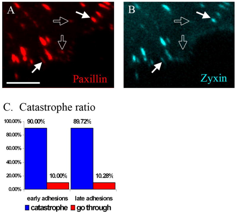
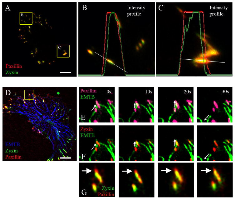
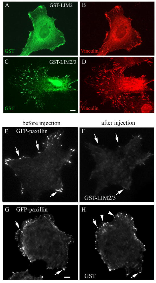
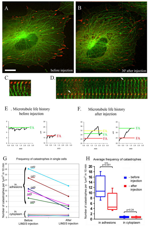
An organized microtubule array is essential for the polarized motility of fibroblasts. Dynamic microtubules closely interact with focal adhesion sites in migrating cells. Here, we examined the effect of focal adhesions on microtubule dynamics. We observed that the probability of microtubule catastrophes (transitions from growth to shrinkage) was seven times higher at focal adhesions than elsewhere. Analysis of the dependence between the microtubule growth rate and catastrophe probability throughout the cytoplasm revealed that a nonspecific (mechanical or spatial) factor provided a minor contribution to the catastrophe induction by decreasing microtubule growth rate at adhesions. Strikingly, at the same growth rate, the probability of catastrophes was significantly higher at adhesions than elsewhere, indicative of a site-specific biochemical trigger. The observed catastrophe induction occurred at adhesion domains containing the scaffolding protein paxillin that has been shown previously to interact with tubulin. Furthermore, replacement of full-length paxillin at adhesion sites by microinjected paxillin LIM2-LIM3 domains suppressed microtubule catastrophes exclusively at adhesions. We suggest that paxillin influences microtubule dynamics at focal adhesions by serving as a scaffold for a putative catastrophe factor and/or regulating its exposure to microtubules.


Figures

Fig. 1
Fig. 1. Microtubule catastrophes at focal adhesions (FA) are specific events
A-B. Kymographs of microtubule dynamics at focal adhesion (A) and in adhesion-free cytoplasm (B). Upper panels show 3xGFP-EMTB (green), mCherry-paxillin (red), microtubule life history plot (white line), catastrophes at adhesion (arrows) catastrophe in adhesion-free area (arrowhead). Lower panels, microtubule images only. Microtubule shrinkage of 0.5 μm or more is considered as a catastrophe. C. A frame from TIRF video sequence of fish fibroblast cell co-transfected with 3xGFPEMTB (green) to visualize microtubules and mCherry-paxillin (red) to mark FA. Bar is 10μm. Boxed region is presented in kymograph in A. D. Microtubule catastrophe distribution in the ventral cell layer. Total of 292 catastrophes in 5 3xGFP-EMTB and mCherry-paxillin co-transfected cells within 12 minutes quantified. E. Average microtubule elongation per catastrophe is 7 times shorter at focal adhesions (blue, 0.71 μm/catastrophe) that elsewhere (red, 4.91 μm/catastrophe). Total of 24 microtubules in 5 3xGFP-EMTB and mCherry-paxillin cells within 12 minutes were quantified.
Fig. 2
Fig. 2. Microtubule tips dynamics at focal adhesions
A. Kymograph of a microtubule catastrophe event at focal adhesion (FA). Microtubule marked with mCherry-EB3 (red), focal adhesion marked with GFP-paxillin (green). B. Kymograph of a microtubule tip (mCherry-EB3, red) which does not undergo catastrophe at the adhesion (GFP-paxillin, green). Microtubule tip changes its growth dynamic at focal adhesion. C. An example of assigned area zones for microtubule growth path near focal adhesion (GFP-paxillin, green). Cytoplasmic zones close to the cell center encoded as zones -2 and -1. Zone adjacent to focal adhesion is encoded as zone 0. Focal adhesion contains zones 1–3, according to its length. Cytoplasmic zones towards the cell periphery are encoded as zones 1out and 2out. Each zone is 1 μm in size. D. Distribution between fast (growth rate >0.1 μm/s) and slow (growth rate <0.1 μm/s) microtubules for each zone. In cytoplasm (yellow background) ~ 70% of microtubules grow with the speed 0.1μm/second or faster. At focal adhesion (green background) percentage of fast microtubule reduces down to ~50%. E. Dependence of microtubule catastrophe ratio (percent of approaching microtubules undergo catastrophe) of time microtubule spends in cytoplasm zone (zone -1, green), at adhesion base (zone 0, blue) and in adhesion (zone 1, red). F. Total microtubule catastrophe ratio for each zone. Percent of approaching microtubules undergo catastrophe increases from 2% in cytoplasm to 25% at focal adhesion. Total of 139 microtubules in 3 3xGFP-EMTB and mCherry-paxillin cells within 15 minutes were quantified. Scale bar 1μm.
Fig. 3
Fig. 3. Microtubule catastrophes do not depend on maturation stage of focal adhesion
A.-B. A frame from video sequence of cell co-transfected with mCherry-paxillin (A. red) and Cerulean-Zyxin (B. cyan). Paxillin marks both early (hollow arrows) and late (filled arrows) focal adhesions. Zyxin is marker for late focal adhesions only (filled arrows). Scale bar 5 μm. C. Microtubule catastrophe ratio. ~90% of approaching microtubules undergo catastrophe both at early and late adhesions. Total 117 catastrophe events in 4 cells were quantified.
Fig. 4
Fig. 4. Catastrophes can occur in the zyxin free zone of focal adhesions
A. A frame from a video sequence of a cell transfected with mCherry-paxillin (red) and GFP-zyxin (green). Scale bar 10μm. B-C. Enlarge areas at the leading edge (B) and trailing edge (C) of the cell (paxillin – red, zyxin – green). The intensity profiles of paxillin (red lines) and zyxin (green lines) along a lines (white) one-pixel wide are shown. Paxillin extends more distantly towards the cell edge. A representative example from 15 cells. D. A frame from a video sequence of a cell transfected with GFP-EMTB (blue) to visualize microtubules, Cerulean-zyxin (green) to mark late adhesions and mCherry-paxillin (red) to mark both early and late adhesions. Scale bar 10μm. Box in enlarged to the right (E-F). E. Enlarged frame sequence of microtubule (green) and paxillin (pink). F. Enlarged frame sequence of microtubule (green) and zyxin (red). Catastrophe happened at time point 20s at the distal zyxin-free end of focal adhesion. Time, seconds. Arrows show direction of microtubule movement. G. Enlarged frame sequence of focal adhesion from 4E,F. Zyxin (green), paxillin (red). Arrows point at zyxin-free distal end of focal adhesion.
Fig. 5
Fig. 5. LIM2/3 displaces full length GFP-paxillin from focal adhesions
Cells microinjected with GST-LIM2 (A,B) and GST-LIM2/3 (C,D) proteins were stained with anti-GST (A,C) and anti-vinculin (B,D) antibodies. Only GST-LIM2/3 (C,D) localizes to focal adhesions. Scale bar 10 μm. Cells expressing GFP-Paxillin (E,G) were microinjected with either GST-LIM2/3 (F) or GST only (H). Within 45 minutes after injection of GST-LIM2/3 paxillin is displaced from existing focal adhesions (F, arrows) and do not appears in new adhesion sites. Microinjection of GST only changed neither paxillin intensity level in old adhesions (H, arrows) nor its ability to incorporate into new adhesion sites (H, arrowheads).
Fig 6
Fig 6. GST-LIM2/3 inhibits microtubule catastrophe at focal adhesions
A-B. Cell expressing GFP-beta-tubulin (green) and mRFP-Zyxin (red) before (A) and after (B) microinjection of GST-LIM2/3 protein. After injection, microtubule density increases as a result of catastrophe suppression at adhesion sites. Scale bar 10 μm. C–D. Kymographs of microtubule dynamics at focal adhesions. Microtubule undergoes catastrophe at adhesion before injection (C) but grows through adhesions after injection (D, arrows). E–F. Examples of microtubule life history plots in cells before (E) and 30 minutes after (F) injecting GST-LIM2/3 construct. Black line indicates tracking position of individual microtubule tips in relation to focal adhesions (FA) marked with red, green and yellow lines. Note multiple catastrophes at adhesions in (E) while microtubules grow through several adhesions without catastrophe in (F). G. Frequency of catastrophes per 1μm2 before and after microinjection of GST-LIM2/3 protein. Upon injection, frequency of catastrophes reduces in focal adhesions (upper bracket) but stays the same in cytoplasm (lower bracket). Data for 271 catastrophes in 5 individual cells recorded both prior to injection and 30 minutes after injection are shown. H. The difference in microtubule catastrophe before (blue boxes) and after (red boxes) injection of GST-LIM2/3 shown in box-and-whisker plot. The P-value was calculated by t-test (Microsoft Excel). Frequency of catastrophes at focal adhesions statistically significantly reduced (p-value <0.001) after injection GST-LIM2/3. At the same time frequency of catastrophes in cytoplasm statistically did not change (p-value=0.24).
Fig 7
Fig 7. Model of paxillin involvement in microtubule catastrophe induction at adhesion sites
In control cells: 1) Paxillin (light blue) binds to adhesion sites (blue) through LIM2/3 domains. Catastrophe factor (Cat F, red box) binds to paxillin through sites other than LIM2/3 domains and in this way enriched at adhesion site. 2A) When microtubule (green) approaches focal adhesion catastrophe factor associated with paxillin induces microtubule catastrophe (red star) and depolymerization (green arrow). Alternatively 2B) When microtubule (green) approaches focal adhesion catastrophe factor associated with paxillin activates microtubule-associated catastrophe-inducing factor (CIF, magenta box) to induce microtubule catastrophe (magenta star) and depolymerization (green arrow). In LIM2/3 injected cells: 3) Exogenous LIM2/3 (light blue) binds to adhesion site and replaces full-length paxillin. Catastrophe factor cannot bind to LIM2/3 mutant and is excluded from adhesion sites. 4) When microtubule approaches focal adhesion it does not undergo catastrophe and continues to polymerize (green arrow) because catastrophe factor is absent or not activated at adhesion.

References

    1. Amos LA, Schlieper D. Microtubules and maps. Adv Protein Chem. 2005;71:257–98. - PubMed
    1. Andrews PD, Ovechkina Y, Morrice N, Wagenbach M, Duncan K, Wordeman L, Swedlow JR. Aurora B regulates MCAK at the mitotic centromere. Dev Cell. 2004;6:253–68. - PubMed
    1. Axelrod D. Total internal reflection fluorescence microscopy. Methods Cell Biol. 1989;30 :245–70. - PubMed
    1. Bershadsky AD, Balaban NQ, Geiger B. Adhesion-dependent cell mechanosensitivity. Annu Rev Cell Dev Biol. 2003;19:677–95. - PubMed
    1. Brown MC, Perrotta JA, Turner CE. Identification of LIM3 as the principal determinant of paxillin focal adhesion localization and characterization of a novel motif on paxillin directing vinculin and focal adhesion kinase binding. J Cell Biol. 1996;135:1109–23. - PMC - PubMed

Publication types