Comparative Study
. 2008 May;149(5):2369-82.
doi: 10.1210/en.2007-0922. Epub 2008 Jan 10.

Trauma and hemorrhage-induced acute hepatic insulin resistance: dominant role of tumor necrosis factor-alpha

Affiliations
Comparative Study

Trauma and hemorrhage-induced acute hepatic insulin resistance: dominant role of tumor necrosis factor-alpha

Jie Xu et al. Endocrinology. 2008 May.

Abstract

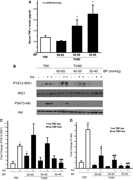
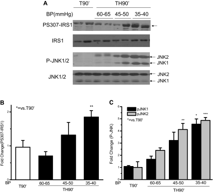
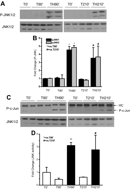
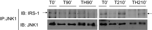
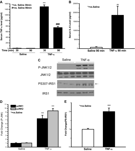
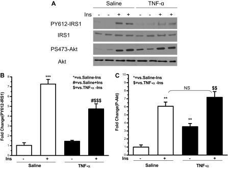
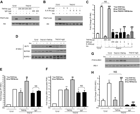
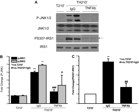
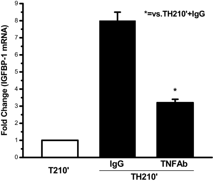
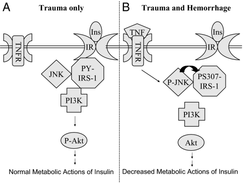
It has long been known that injury, infections, and other critical illnesses are often associated with hyperglycemia and hyperinsulinemia. Mortality of critically ill patients is greatly reduced by intensive insulin therapy, suggesting the significance of reversing or compensating for the development of acute insulin resistance. However, the development of acute injury/infection-induced insulin resistance is poorly studied, much less than the chronic diseases associated with insulin resistance, such as type 2 diabetes and obesity. We previously found that insulin resistance develops acutely in the liver after trauma and hemorrhage. The present study was designed to begin to understand the first steps in the development of trauma and hemorrhage-induced acute hepatic insulin resistance in an animal model of injury and blood loss similar to traumatic or surgical injury and hemorrhage. We present novel data that indicate that hepatic insulin resistance increased dramatically with an increasing extent of hemorrhage. With increasing extent of blood loss, there were increases in serum TNF-alpha levels, phosphorylation of liver insulin receptor substrate-1 on serine 307, and liver c-Jun N-terminal kinase activation/phosphorylation. Exogenous TNF-alpha infusion increased c-Jun N-terminal kinase phosphorylation and insulin receptor substrate-1 serine 307 phosphorylation, and inhibited insulin-induced signaling in liver. Conversely, neutralizing TNF-alpha antibody treatment reversed many of the hemorrhage-induced changes in hepatic insulin signaling. Our data indicate that the acute development of insulin resistance after trauma and hemorrhage may have some similarities to the insulin resistance that occurs in chronic diseases. However, because so little is known about this acute insulin-resistant state, much more needs to be done before we can attain a level of understanding similar to that of chronic states of insulin resistance.


Figures

Figure 1
Figure 1
Correlation between serum TNF-α levels, the inhibition of hepatic insulin (Ins) signaling, and the extent of hemorrhage after trauma alone or trauma and hemorrhage. Rats were subjected to T or trauma and hemorrhage. In the trauma and hemorrhage group, rats were bled to a mean arterial pressure of 60–65, 45–50, or 35–40 mm Hg within 10 min and then maintained for 90 min. A, Serum TNF-α levels. Data are presented as the mean + sem of samples from four to eight rats in each group. B, Saline (−) or 5 U insulin (+) was injected into the portal vein, and 1 min later the liver was removed, and protein extracts were prepared, resolved by SDS-PAGE, and subjected to Western blot analysis using specific anti-PY612-IRS1, anti-IRS1, anti-PS473-Akt, and anti-Akt antibodies. Representative Western blots are presented. The original blots were cut and rearranged for the sake of clarity. C and D, Autoradiographs were quantified by scanning densitometry. Data are presented as the mean ± sem of samples from four to eight rats in each group. In this and all the following figures: * or #, P < 0.05; ** or ##, P < 0.01; and *** or ###, P < 0.001. BP, Blood pressure.
Figure 2
Figure 2
Serine 307 phosphorylation of IRS1 and phosphorylation of JNK1/2 were correlated with the extent of hemorrhage in rat liver after trauma and hemorrhage. At the same time points and treatment regimens described in Fig. 1, the liver was removed, and protein extracts were subjected to Western blot analysis with specific anti-PS307-IRS1, anti-IRS1, anti-P-JNK1/2, and anti-JNK1/2 antibodies. A, Representative Western blots are presented. B and C, Data are presented as the mean ± sem of samples from four to eight rats in each group. BP, Blood pressure.
Figure 3
Figure 3
Phosphorylation and activity of JNK1/2 increased in rat liver after trauma and hemorrhage. At 0′ (after T, but before any further treatment), 90′ (without or with hemorrhage), or 210′ (without hemorrhage or with hemorrhage and 60 min recovery after 60 min resuscitation), the liver was removed. A, Protein extracts were subjected to Western blot analysis with specific anti-P-JNK1/2 and anti-JNK1/2 antibodies. Representative Western blots are presented. C, Protein extracts were subjected to in vitro JNK kinase activity assay using c-Jun as the substrate. Representative Western blots are presented. HC indicates the heavy chain of immunoprecipitated anti-JNK antibody. B and D, Data are presented as the mean ± sem of samples from four to eight rats in each group.
Figure 4
Figure 4
The interaction between JNK and IRS1 in rat liver. At the same time points and treatment regimens described in Fig. 3, the liver was removed, and protein extracts were immunoprecipitated with specific anti-JNK1 antibody and then subjected to Western blot analysis by specific anti-IRS-1 and anti-JNK antibodies. Representative Western blots are presented. IP, immunoprecipitation; IB, immunoblot.
Figure 5
Figure 5
Exogenous TNF-α infusion induced JNK phosphorylation and IRS-1 S307 phosphorylation in rat liver. TNF-α at a dose of 125 μg/kg body weight (in 1 ml saline containing 0.2% BSA) or 1 ml saline containing 0.2% BSA was infused via the portal vein over a period of 30 min at a constant infusion rate. A and B, Blood was collected at 30 min of TNF-α or saline infusion, or at 60 min after a 30-min infusion. Serum TNF-α and IL-6 levels were examined. C, Sixty minutes after 30 min of TNF-α or saline infusion, either saline or insulin was injected into the portal vein, and 1 min later the liver was removed. Liver protein was subjected to Western blot analysis with specific anti-P-JNK, anti-JNK, anti-PS307-IRS-1, and anti-IRS-1 antibodies. Representative Western blots are presented. D and E, Data are presented as the mean ± sem of samples from three rats in each group.
Figure 6
Figure 6
Insulin (Ins)-induced hepatic tyrosine phosphorylation of IRS-1 and phosphorylation of Akt were inhibited by exogenous TNF-α infusion. At the same time points and treatment regimens described in Fig. 5, the liver was removed, and protein extracts were subjected to Western blot analysis by specific anti-PY612-IRS-1, anti-IRS-1, anti-PS473-Akt, and anti-Akt antibodies. A, Representative Western blots are presented. B and C, Data are presented as the mean ± sem of samples from three rats in each group. $, P < 0.05. $$, P < 0.01. $$$, P < 0.001. NS, Not significant.
Figure 7
Figure 7
Neutralizing TNF-α antibody restored insulin (Ins)-induced phosphorylation of Akt, tyrosine phosphorylation of IRS-1, and IRS-1/PI3K association after trauma and hemorrhage. Neutralizing TNF-α antibody (200 μg), IL-6 antibody (5 μg), or IgG (200 or 5 μg) was administered as described in Materials and Methods. After the completion of infusion, either saline or insulin was injected into the portal vein, and 1 min later the liver was removed. A and B, Liver protein was subjected to Western blot analysis with specific anti-P-Akt, and anti-Akt antibodies. Representative Western blots are presented. D, Liver protein extracts were immunoprecipitated with specific anti-IRS-1 antibody and then subjected to Western blot analysis by specific anti-phospho-tyrosine (PY), anti-PI3K, and anti-IRS-1 antibodies. G, Liver protein was subjected to Western blot analysis with specific anti-PY612-IRS-1 and anti-IRS-1 antibodies. Representative Western blots are presented. C, E, F, and H, Data are presented as the mean ± sem of samples from three to four rats in each group. NS, Not significant; IP, immunoprecipitation; IB, immunoblot.
Figure 8
Figure 8
Trauma and hemorrhage-induced phosphorylation of JNK and S307 phosphorylation of IRS-1 was inhibited by neutralizing TNF-α antibody. At the same time points and treatment regimens described in Fig. 7, the liver was removed, and protein extracts were subjected to Western blot analysis by specific anti-P-JNK, anti-JNK, anti-PS307-IRS-1, and anti-IRS-1 antibodies. A, Representative Western blots are presented. B and C, Data are presented as the mean ± sem of samples from three rats in each group.
Figure 9
Figure 9
Neutralizing TNF-α antibody restored insulin’s inhibition of IGFBP-1 mRNA expression after trauma and hemorrhage. In the T210′ group and the TH210′ group pretreated with TNF-α antibody or IgG, the liver was removed, and total RNA was isolated and subjected to Northern analysis using an IGFBP-1 probe. The data are presented as the mean + sem of samples from three rats in each group.
Figure 10
Figure 10
Proposed model for TNF-α-mediated acute hepatic insulin (Ins) resistance after trauma and hemorrhage. A, In the liver of trauma-only animals, insulin binds to IR on the cell membrane, resulting in tyrosine phosphorylation of IRS-1, association of PI3K with IRS-1, and Akt phosphorylation. JNK is associated with IRS-1, but JNK is not phosphorylated or activated. B, In the liver of trauma and hemorrhage animals, local TNF-α levels rapidly increase and bind to the TNF receptor (TNFR) on the cell membrane, leading to phosphorylation and activation of JNK. Activated JNK in turn induces S307 phosphorylation of IRS-1 and inhibits tyrosine phosphorylation of IRS-1. This leads to a loss of association of PI3K with IRS-1 and impaired Akt phosphorylation.

Similar articles

Cited by

References

    1. Del Aguila LF, Krishnan RK, Ulbrecht JS, Farrell PA, Correll PH, Lang, CH, Zierath JR, Kirwan JP 2000 Muscle damage impairs insulin stimulation of IRS-1, PI 3-kinase, and Akt-kinase in human skeletal muscle. Am J Physiol Endocrinol Metab 279:E206–E212 - PubMed
    1. Chaudry IH, Sayeed MM, Baue AE 1974 Insulin resistance in experimental shock. Arch Surg 109:412–415 - PubMed
    1. Ikezu T, Okamoto T, Yonezawa K, Tompkins RG, Martyn JA 1997 Analysis of thermal injury-induced insulin resistance in rodents. Implication of postreceptor mechanisms. J Biol Chem 272:25289–25295 - PubMed
    1. Carter EA 1998 Insulin resistance in burns and trauma. Nutr Rev 56(1 Pt 2):S170–S176 - PubMed
    1. Van den Berghe G, Wouters P, Weekers F, Verwaest C, Bruyninckx F, Schetz M, Vlasselaers D, Ferdinande P, Lauwers P, Bouillon R 2001 Intensive insulin therapy in the critically ill patients. N Engl J Med 345:1359–1367 - PubMed

Publication types

MeSH terms

Substances