. 2008 Jan 23;27(2):394-405.
doi: 10.1038/sj.emboj.7601966. Epub 2008 Jan 10.

SAGA-mediated H2B deubiquitination controls the development of neuronal connectivity in the Drosophila visual system

Affiliations

SAGA-mediated H2B deubiquitination controls the development of neuronal connectivity in the Drosophila visual system

Vikki M Weake et al. EMBO J. .

Abstract

Nonstop, which has previously been shown to have homology to ubiquitin proteases, is required for proper termination of axons R1-R6 in the optic lobe of the developing Drosophila eye. Herein, we establish that Nonstop actually functions as an ubiquitin protease to control the levels of ubiquitinated histone H2B in flies. We further establish that Nonstop is the functional homolog of yeast Ubp8, and can substitute for Ubp8 function in yeast cells. In yeast, Ubp8 activity requires Sgf11. We show that in Drosophila, loss of Sgf11 function causes similar photoreceptor axon-targeting defects as loss of Nonstop. Ubp8 and Sgf11 are components of the yeast SAGA complex, suggesting that Nonstop function might be mediated through the Drosophila SAGA complex. Indeed, we find that Nonstop does associate with SAGA components in flies, and mutants in other SAGA subunits display nonstop phenotypes, indicating that SAGA complex is required for accurate axon guidance in the optic lobe. Candidate genes regulated by SAGA that may be required for correct axon targeting were identified by microarray analysis of gene expression in SAGA mutants.


Figures

Figure 1
Figure 1
Orthologs of the proteins required for H2B deubiquitination in yeast were identified in Drosophila. (A) Alignment of D. melanogaster Nonstop and S. cerevisiae Ubp8. The conserved zinc finger, Cys-box and His-box domains are indicated. (B) Dendrogram of the 16 yeast and 18 Drosophila ubiquitin proteases. The yeast Ubps are indicated in italics. Nonstop and Ubp8 form a single clade (box). (C) Alignment of D. melanogaster Sgf11/CG13379 and S. cerevisiae Sgf11. Key cysteine and histidine residues are indicated by asterisks. (D) Dendrogram showing the relationship between human ATAXIN, S. cerevisiae Sgf11 and Sgf73, S. pombe Sgf11 and D. melanogaster CG13379/Sgf11. (E) Four-fold serial dilutions of ubp8Δgcn5Δ yeast were grown at 30°C for 2 days on galactose plates. The growth defect can be rescued by the introduction of yeast UBP8 or Drosophila Nonstop.
Figure 2
Figure 2
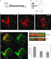
Axon targeting is disrupted in the sgf11 mutant. (A) Schematic representation of the sgf11 (CG13379) locus, showing the position of the piggyBac E01308 transposon (insertion). The single exon is represented by a box. Translated sequences are filled with gray, and 5′ and 3′ untranslated regions are open boxes. (B) RT–PCR from WT (lanes 1 and 4) or sgf11 (lanes 2 and 5) third instar larvae with primers specific for the constitutively expressed gene RpL32 (lanes 1–3) or for sgf11 (lanes 4–6). Lanes 3 and 6 correspond to the negative PCR control. (CF) In third instar larvae, photoreceptor cells from the eye disc extend axons through the optic stalk (os) into the optic lobe. R1–R6 axons terminate in the lamina (la) in wild type, while R7–R8 project through the lamina into the medulla (me). The projection pattern of R2–R5 was visualized in wild type (C), sgf11 (D) and nonstop (E) larval optic lobes using the ro-τlacZ marker (red). In wild-type larvae, R2–R5 axons terminate in the lamina. However, in nonstop and sgf11 larvae, many R2–R5 axons fail to terminate in the lamina (arrowheads), and instead project into the medulla. (F) A triple layer of glial cells, visualized using anti-repo (green), is present at the lamina in wild type (dotted lines) where R2–R5 growth cones terminate. These glial cells migrate from regions at the dorsal and ventral margins of the lamina (arrowheads) into the lamina. In the sgf11 mutant, an increased number of glial cells accumulate at the edges of the lamina (arrowheads) and fewer glial cells are present along the lamina. (C–F) Scale bars, 20 μm. (G, H) The number of repo-positive glial cells (green) along a given length of the lamina (ro-τlacZ, red) was compared in wild-type, nonstop and sgf11 mutants.
Figure 3
Figure 3
Nonstop and Sgf11 associate and are both required for H2B deubiquitination and regulation of gene expression. (A) Extract from S2 cells transfected with pRmHa3-Nonstop-HA2FL2 and pMT-Sgf11-V5 or untransfected S2 cells (−) was incubated with FLAG-M2-agarose beads. The immunoprecipitated material (IP, lanes 3 and 4) was analyzed by western blotting relative to 4% input (lanes 1 and 2). (B) Monoubiquitinated H2B levels increase in the nonstop and sgf11 mutants. Histones were acid-extracted from OregonR (WT), nonstop or sgf11 third instar larvae nuclei and analyzed by western blotting using antibodies against H2B. Mean results from three separate experiments are graphed normalized to H2B levels. (C) Nonstop and Sgf11 coregulate a large subset of genes. The number of overlapping genes with increased or decreased transcript levels greater than two-fold in nonstop and sgf11 homozygous mutant third instar larvae compared to their heterozygote siblings (P<0.05 for ⩾2 biological replicates).
Figure 4
Figure 4
Mutations in nonstop and sgf11 do not affect H3K9 acetylation and have distinct but overlapping effects on gene expression when compared to a dSAGA mutation. (A) Histones were acid-extracted from OregonR (WT), nonstop, sgf11 or ada2b third instar larvae nuclei and analyzed by western blotting using antibodies against H2B and acetylated H3 Lys-9. Mean results from three separate experiments are graphed normalized to H2B levels. (B) The number of overlapping genes with increased or decreased transcript levels greater than two-fold in nonstop, sgf11 and ada2b homozygous mutant third instar larvae compared to their heterozygote siblings (P<0.05 for ⩾2 biological replicates).
Figure 5
Figure 5
Nonstop and Sgf11 are associated with dSAGA. (A) MudPIT analysis of dSAGA, affinity purified from cells expressing tagged dAda1, WDA or dAda2b identified Nonstop and CG13379/Sgf11 as putative components of dSAGA. The table shows the number of non-redundant spectra for each protein (total peptides) and the amino-acid sequence coverage (% coverage). (B) Extract from S2 cells transfected with pRmHa3-Nonstop-HA2FL2 was immunoprecipitated as described in Figure 3. Immunoprecipitated material (IP, lanes 3 and 4) was analyzed by western blotting relative to 1% input (lanes 1 and 2). (C) Extract from S2 cells transfected with pRmHa3-Sgf11-HA2FL2 was incubated with HA-agarose beads, and the immunoprecipitated material analyzed as in (B). (D) Sgf11-HA2FL2 Ni-agarose HAT-enriched S2 cell nuclear extract was applied to a Mono-Q column and the elution profiles of dGcn5 (dKAT2), Ada2b, Spt3 and Sgf11 compared by western blotting. I, input; F, unbound; Ni, eluted from Ni-agarose. (E) Antibodies against dGcn5 (dKAT2) were added to 1 mg of Nonstop-HA2FL2 extract. Equal amounts of input (I), mock depletion (−) and immunodepleted extract (+, ++) were analyzed by western blotting for the presence of dGcn5 (dKAT2), Nonstop and tubulin.
Figure 6
Figure 6
Axon targeting is disrupted in the ada2b mutant. (AE) The R-cell projection pattern in eye–brain complexes from wild-type and ada2b larvae were examined using mAB24B10 (R1–R8, red; A, C) or ro-τlacZ (R2–R5; B, D, E) relative to the glial cell position (anti-repo, green; A, C, E). A single plane is shown in A–D, and 3D reconstructed images in E. In the wild type, R1–R6 axons terminate in the lamina (la) between rows of epithelial glial cells (eg), and marginal (mg) and medulla (meg) glial cells. R7–R8 project through the lamina into the medulla (me). In the ada2b mutant, some R1–R6 cells project inappropriately into the medulla (arrowheads), and there are gaps in the lamina plexus accompanied by a reduction in glial cell number. (E) More glial cells are present at the ventral and dorsal margins (arrows) of the lamina (dotted lines) in ada2b relative to the wild type. Two different images representing the variability in axon mistargeting in ada2b are shown in (E). Scale bars, 20 μm. (F) The number of glial cells along a given length of the lamina was compared in wild type and ada2b as described in Figure 2. (G) Extract from UAS-Ada2b-HAFL2/gcm-GAL4 larval CNS/eye-antennal disc complexes was incubated with HA-agarose beads. Immunoprecipitated material (IP, lane 2) was analyzed relative to 1% input (lane 1) by western blotting.

References

    1. Allis CD, Berger SL, Cote J, Dent S, Jenuwien T, Kouzarides T, Pillus L, Reinberg D, Shi Y, Shiekhattar R, Shilatifard A, Workman J, Zhang Y (2007) New nomenclature for chromatin-modifying enzymes. Cell 131: 633–636 - PubMed
    1. Awasaki T, Ito K (2004) Engulfing action of glial cells is required for programmed axon pruning during Drosophila metamorphosis. Curr Biol 14: 668–677 - PubMed
    1. Brennan CA, Li TR, Bender M, Hsiung F, Moses K (2001) Broad-complex, but not ecdysone receptor, is required for progression of the morphogenetic furrow in the Drosophila eye. Development 128: 1–11 - PubMed
    1. Carre C, Szymczak D, Pidoux J, Antoniewski C (2005) The histone H3 acetylase dGcn5 is a key player in Drosophila melanogaster metamorphosis. Mol Cell Biol 25: 8228–8238 - PMC - PubMed
    1. Chen X, Fischer JA (2000) In vivo structure/function analysis of the Drosophila fat facets deubiquitinating enzyme gene. Genetics 156: 1829–1836 - PMC - PubMed

Publication types

MeSH terms