

The biomechanics of amnion rupture: an X-ray diffraction study
- PMID: 18188405
- PMCID: PMC2190618
- DOI: 10.1371/journal.pone.0001147
The biomechanics of amnion rupture: an X-ray diffraction study
Abstract
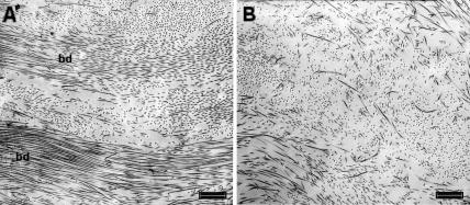
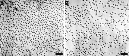
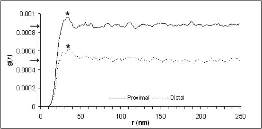
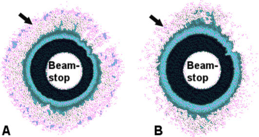
Pre-term birth is the leading cause of perinatal and neonatal mortality, 40% of which are attributed to the pre-term premature rupture of amnion. Rupture of amnion is thought to be associated with a corresponding decrease in the extracellular collagen content and/or increase in collagenase activity. However, there is very little information concerning the detailed organisation of fibrillar collagen in amnion and how this might influence rupture. Here we identify a loss of lattice like arrangement in collagen organisation from areas near to the rupture site, and present a 9% increase in fibril spacing and a 50% decrease in fibrillar organisation using quantitative measurements gained by transmission electron microscopy and the novel application of synchrotron X-ray diffraction. These data provide an accurate insight into the biomechanical process of amnion rupture and highlight X-ray diffraction as a new and powerful tool in our understanding of this process.
Conflict of interest statement
Figures
References
-
- Bourne GL. The microscopic anatomy of the human amnion and chorion. Am J Obstet Gynecol. 1960;79:1070–1073. - PubMed
-
- Keirse MJ. An evaluation of formal risk scoring for preterm birth. Am J Perinatol. 1989;6:226–233. - PubMed
-
- Goldenberg RL, Rouse DJ. Prevention of Premature Birth. N Engl J Med. 1998;339:313–320. - PubMed
-
- Moore RM, Mansour JM, Redline RW, Mercer BM, Moore JJ. The Physiology of Fetal Membrane Rupture: Insight Gained from the Determination of Physical Properties. Placenta. 2006;27:1037–1051. - PubMed
-
- Malak TM, Ockleford CD, Bell SC, Dalgleish R, Bright N, et al. Confocal immunofluorescence localization of collagen types I, III, IV, V and VI and their ultrastructural organization in term human fetal membranes. Placenta. 1993;14:385–406. - PubMed
Publication types
MeSH terms
Grants and funding
LinkOut - more resources
Full Text Sources
Medical
