. 2008 Feb;118(2):619-28.
doi: 10.1172/JCI33970.

Claudin-16 and claudin-19 interact and form a cation-selective tight junction complex

Affiliations

Claudin-16 and claudin-19 interact and form a cation-selective tight junction complex

Jianghui Hou et al. J Clin Invest. 2008 Feb.

Abstract

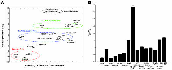
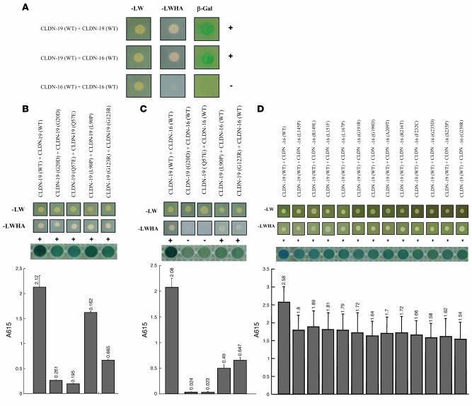
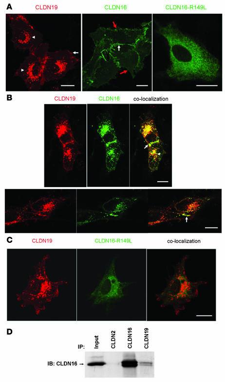
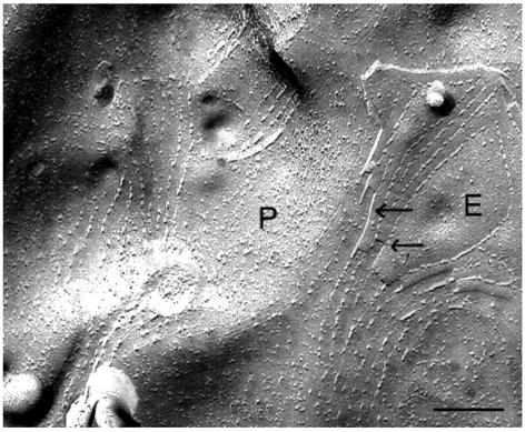
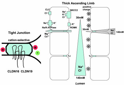
Tight junctions (TJs) play a key role in mediating paracellular ion reabsorption in the kidney. Familial hypomagnesemia with hypercalciuria and nephrocalcinosis (FHHNC) is an inherited disorder caused by mutations in the genes encoding the TJ proteins claudin-16 (CLDN16) and CLDN19; however, the mechanisms underlying the roles of these claudins in mediating paracellular ion reabsorption in the kidney are not understood. Here we showed that in pig kidney epithelial cells, CLDN19 functioned as a Cl(-) blocker, whereas CLDN16 functioned as a Na(+) channel. Mutant forms of CLDN19 that are associated with FHHNC were unable to block Cl(-) permeation. Coexpression of CLDN16 and CLDN19 generated cation selectivity of the TJ in a synergistic manner, and CLDN16 and CLDN19 were observed to interact using several criteria. In addition, disruption of this interaction by introduction of FHHNC-causing mutant forms of either CLDN16 or CLDN19 abolished their synergistic effect. Our data show that CLDN16 interacts with CLDN19 and that their association confers a TJ with cation selectivity, suggesting a mechanism for the role of mutant forms of CLDN16 and CLDN19 in the development of FHHNC.


Figures

Figure 1
Figure 1. Effects of CLDN16, CLDN19, and their mutants in LLC-PK1 cells on paracellular ion conductance.
Dilution potential values (A) and PNa/PCl (B) across LLC-PK1 cell monolayers expressing CLDN16, CLDN19, and their mutants, individually or in pairs, are shown.
Figure 2
Figure 2. CLDN16 interacts with CLDN19 in yeast.
(A) Y2H assays showing interaction of CLDN16-WT with CLDN19-WT and CLDN19-WT with CLDN19-WT, but not of CLDN16-WT with CLDN16-WT. Shown are plates with selective medium lacking leucine and tryptophan (–LW), indicating the transforming of both bait and prey vectors; with SD-LWHA, indicating the expression of reporter genes HIS3 and ADE2; and β-galactosidase assay (A615 values) for quantification of interaction strength. (B) Mutations in CLDN19 affecting its homomeric interaction. (C) Mutations in CLDN19 affecting its heteromeric interaction with CLDN16. (D) Mutations in CLDN16 affecting its heteromeric interaction with CLDN19.
Figure 3
Figure 3. Cotrafficking and coimmunoprecipitation between CLDN16 and CLDN19 in unpolarized epithelial cells.
(A) Confocal images showing subcellular localization of CLDN19, CLDN16, and CLDN16-R149L mutant in LLC-PK1 cells. CLDN19 localized to the plasma membrane (arrow) and the endosomes and lysosomes (arrowheads), CLDN16 localized to the plasma membrane (red arrows); and CLDN16-R149L was confined to the ER. In the middle panel, the white arrow denotes a site of cell-cell interaction. (B) Coexpression of CLDN16 with CLDN19 in LLC-PK1 cells altered the subcellular localization of CLDN16. Notably, CLDN16 was recruited to the endosomes and lysosomes (arrowhead), where colocalization with CLDN19 occurred. CLDN16 and CLDN19 colocalization was also found at the cell-cell interaction (arrow). (C) The confinement of CLDN16-R149L mutant to the ER was not affected by CLDN19 coexpression. No colocalization was found between CLDN16-R149L and CLDN19. (D) Coimmunoprecipitation of CLDN16 and CLDN19 cotransfected in HEK293 cells. Input lane shows 10% of input amount. Antibodies used for coimmunoprecipitation are shown above the lanes; antibody for blot visualization is shown at left. Scale bars: 10 μm.
Figure 4
Figure 4. Freeze-fracture image of CLDN19-transfected L cells revealed TJ structures.
Notably, a P-to-E fracture face transition was seen (arrows) where there was alignment of the TJ strands. Scale bar: 1 μm.
Figure 5
Figure 5. Freeze-fracture images of CLDN19/CLDN16 cotransfected L cells with immunogold (10 nm gold particle) labeling for CLDN19 and CLDN16.
Note the localization of CLDN19 (A) and CLDN16 (B) within the well-developed networks of TJ strands. Scale bars: 200 nm.
Figure 6
Figure 6. Function of TJ and mechanism of Mg2+ reabsorption.
In TAL, NaCl reabsorption is mediated through Na+K+Cl-cotransporter type 2 (NKCC2) in the apical membrane. On the basolateral side, Na/K-ATPase provides the energy source and also allows Na+ to exit the cell in exchange for K+ entry. K+ is secreted to the lumen side via the renal outer medullary K+ channel (ROMK). Cl exits the cell via the Cl channel (CLC). Continuous NaCl reabsorption along TAL results in gradual tubular fluid dilution and the development of a transepithelial NaCl concentration gradient (from peritubular space, 140 mM, down to lumen, 30 mM). As the epithelial cells in TAL are joined by cation-selective TJs, the NaCl concentration gradient results in a lumen-positive transepithelial diffusion potential. This positive potential drives the reabsorption of Mg2+ and Ca2+ through the TJ (known as the paracellular pathway). CLDN16 and CLDN19 interact in TJs and contribute to the cation selectivity of TJs.

References

    1. Simon D.B., et al. Paracellin-1, a renal tight junction protein required for paracellular Mg2+ resorption. . Science. 1999;285:103–106. - PubMed
    1. Konrad M., et al. 2006Mutations in the tight-junction gene claudin 19 (CLDN19) are associated with renal magnesium wasting, renal failure, and severe ocular involvemen t . Am. J. Hum. Genet. 79949–957. - PMC - PubMed
    1. Anderson J.M., Van Itallie C.M., Fanning A.S. Setting up a selective barrier at the apical junction complex. Curr. Opin. Cell Biol. 2004;16:140–145. - PubMed
    1. Hou J., Paul D.L., Goodenough D.A. Paracellin-1 and the modulation of ion selectivity of tight junctions. J. Cell Sci. 2005;118:5109–5118. - PubMed
    1. Hou J., Gomes A.S., Paul D.L., Goodenough D.A. Study of claudin function by RNA interference. J. Biol. Chem. 2006;281:36117–36123. - PubMed

Publication types