

Review
doi: 10.1007/978-0-387-74904-4_43.
Alphavbeta5 integrin receptors at the apical surface of the RPE: one receptor, two functions
Affiliations
- PMID: 18188966
- PMCID: PMC3630459
- DOI: 10.1007/978-0-387-74904-4_43
Item in Clipboard
Review
Alphavbeta5 integrin receptors at the apical surface of the RPE: one receptor, two functions
Adv Exp Med Biol.
2008.
No abstract available
Figures

Fluorescence microscopy quantification of POS phagocytosis by RPE cells in vitro and in vivo. (a) Maximal projection of confocal microscopy sections of primary wild-type mouse RPE cells in culture. RPE cells show vigorous uptake of FITC-labeled POS (white) after 1 hour of phagocytic challenge. RPE cell junctions stained with ZO-1 (gray outlines). (b–d) Cryosections of 2-month-old wild-type mice eyecups labeled with rhodopsin antibody. (b) RPE-photoreceptor outer segment interface close-up showing intact rod outer segments and opsin-positive phagosomes (bright white dots) adjacent to RPE nuclei (gray). (c–d) Low magnification view of similar stainings without nuclei illustrates that these images can be used to count phagosome numbers in the RPE. (c) One hour before light onset, the RPE cell layer shows few opsin-labeled phagosomes. (d) Two hours after light onset, the RPE cell layer shows numerous opsin-labeled phagosomes confirming the daily burst of rod POS phagocytosis by the RPE. Scale bars: 20 μm. Modified from Finnemann and Chang with permission from Humana Press Inc.
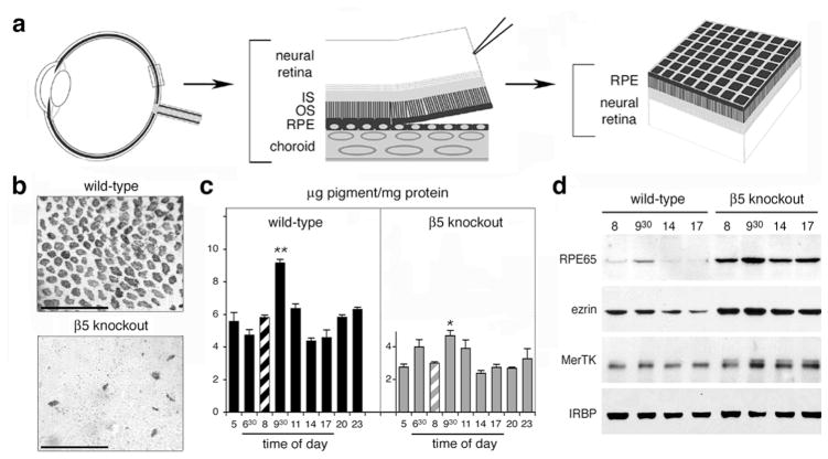
Retinal adhesion is dramatically reduced in β5 knockout mice. (a) After enucleation and lens/cornea removal, retinas are swiftly peeled off opened eyecups creating shearing forces to assess retinal adhesion. In retinas with normal retinal adhesion, apical cellular domains of RPE largely remain attached to the outer surface of the neural retina. (b) Whole-mount of peeled retina, shown outer retina up, demonstrating that β5 knockout retina retains significantly less RPE pigment than wild-type retina. (c) Quantification of RPE pigment in retina peeled at different times of day. β5 knockout retina shows reduced pigment contents and attenuated adhesiveness peak compared to wild-type retina. (d) Representative immunoblots of individual peeled retina confirming the melanin pigment results. Modified from Finnemann and Chang and from Nandrot et al. (2006) with permission from Humana Press Inc. and the American Physiological Society.
Similar articles
-
Integrin alphavbeta5 participates in the binding of photoreceptor rod outer segments during phagocytosis by cultured human retinal pigment epithelium.Invest Ophthalmol Vis Sci. 1998 Aug;39(9):1703-12. Invest Ophthalmol Vis Sci. 1998. PMID: 9699560
-
Essential role for MFG-E8 as ligand for alphavbeta5 integrin in diurnal retinal phagocytosis.Proc Natl Acad Sci U S A. 2007 Jul 17;104(29):12005-10. doi: 10.1073/pnas.0704756104. Epub 2007 Jul 9. Proc Natl Acad Sci U S A. 2007. PMID: 17620600 Free PMC article.
-
Integrin alphavbeta5 is not required for the phagocytosis of photoreceptor outer segments by cultured retinal pigment epithelial cells.Exp Eye Res. 2003 Sep;77(3):281-6. doi: 10.1016/s0014-4835(03)00158-1. Exp Eye Res. 2003. PMID: 12907160
-
Role of alphavbeta5 integrin in regulating phagocytosis by the retinal pigment epithelium.Adv Exp Med Biol. 2003;533:337-42. doi: 10.1007/978-1-4615-0067-4_42. Adv Exp Med Biol. 2003. PMID: 15180282 Review. No abstract available.
-
Morphogenesis of the retinal pigment epithelium: toward understanding retinal degenerative diseases.Ann N Y Acad Sci. 1998 Oct 23;857:1-12. doi: 10.1111/j.1749-6632.1998.tb10102.x. Ann N Y Acad Sci. 1998. PMID: 9917828 Review.
Cited by
-
Genetic dissection of TAM receptor-ligand interaction in retinal pigment epithelial cell phagocytosis.Neuron. 2012 Dec 20;76(6):1123-32. doi: 10.1016/j.neuron.2012.10.015. Neuron. 2012. PMID: 23259948 Free PMC article.
-
Mechanisms of extracellular vesicle uptake in stressed retinal pigment epithelial cell monolayers.Biochim Biophys Acta Mol Basis Dis. 2020 Mar 1;1866(3):165608. doi: 10.1016/j.bbadis.2019.165608. Epub 2019 Nov 15. Biochim Biophys Acta Mol Basis Dis. 2020. PMID: 31740401 Free PMC article.
-
MicroRNA-204/211 alters epithelial physiology.FASEB J. 2010 May;24(5):1552-71. doi: 10.1096/fj.08-125856. Epub 2010 Jan 7. FASEB J. 2010. PMID: 20056717 Free PMC article.
-
Light-responsive microRNA miR-211 targets Ezrin to modulate lysosomal biogenesis and retinal cell clearance.EMBO J. 2020 Apr 15;39(8):e102468. doi: 10.15252/embj.2019102468. Epub 2020 Mar 10. EMBO J. 2020. PMID: 32154600 Free PMC article.
-
Integrins as Therapeutic Targets: Successes and Cancers.Cancers (Basel). 2017 Aug 23;9(9):110. doi: 10.3390/cancers9090110. Cancers (Basel). 2017. PMID: 28832494 Free PMC article. Review.
References
-
- Cook B, Lewis GP, Fisher SK, Adler R. Apoptotic photoreceptor degeneration in experimental retinal detachment. Invest Ophthalmol Vis Sci. 1995;36:990–996. - PubMed
-
- Edwards RB, Szamier RB. Defective phagocytosis of isolated rod outer segments by RCS rat retinal pigment epithelium in culture. Science. 1977;197:1001–1003. - PubMed
-
- Endo EG, Yao XY, Marmor MF. Pigment adherence as a measure of retinal adhesion: dependence on temperature. Invest Ophthalmol Vis Sci. 1988;29:1390–1396. - PubMed
-
- Feeney L. Lipofuscin and melanin of human retinal pigment epithelium. Fluorescence, enzyme cytochemical, and ultrastructural studies. Invest Ophthalmol Vis Sci. 1978;17:583–600. - PubMed
Publication types
MeSH terms
Substances
Grants and funding
LinkOut - more resources
Full Text Sources
Other Literature Sources
