. 2008 Jan 11:5:6.
doi: 10.1186/1743-422X-5-6.

Down-regulation of cell surface CXCR4 by HIV-1

Affiliations

Down-regulation of cell surface CXCR4 by HIV-1

Bongkun Choi et al. Virol J. .

Abstract

Background: CXC chemokine receptor 4 (CXCR4), a member of the G-protein-coupled chemokine receptor family, can serve as a co-receptor along with CD4 for entry into the cell of T-cell tropic X4 human immunodeficiency virus type 1 (HIV-1) strains. Productive infection of T-lymphoblastoid cells by X4 HIV-1 markedly reduces cell-surface expression of CD4, but whether or not the co-receptor CXCR4 is down-regulated has not been conclusively determined.

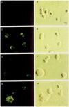
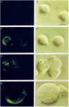
Results: Infection of human T-lymphoblastoid cell line RH9 with HIV-1 resulted in down-regulation of cell surface CXCR4 expression. Down-regulation of surface CXCR4 correlated temporally with the increase in HIV-1 protein expression. CXCR4 was concentrated in intracellular compartments in H9 cells after HIV-1 infection. Immunofluorescence microscopy studies showed that CXCR4 and HIV-1 glycoproteins were co-localized in HIV infected cells. Inducible expression of HIV-1 envelope glycoproteins also resulted in down-regulation of CXCR4 from the cell surface.

Conclusion: These results indicated that cell surface CXCR4 was reduced in HIV-1 infected cells, whereas expression of another membrane antigen, CD3, was unaffected. CXCR4 down-regulation may be due to intracellular sequestering of HIV glycoprotein/CXCR4 complexes.


Figures

Figure 1
Figure 1
Flow cytometry analysis demonstrating reduced CXCR4 expression in HIV-1 infected RH9 cells. Panel A: RH9 T-lymphoblastoid cells infected with HIV-1LA1. On days 1, 4, and 7 postinfection cells were fixed with 4% paraformaldehyde, stained with mouse MAb 12G5 anti-CXCR4 (10 μg/ml) or isotype-matched control antibody followed by fluorescein isothiocyanate (FITC)-conjugated goat anti-mouse immunoglobulin G, and analyzed by flow cytometry. Median fluorescence intensity was calculated as an indicator of the level of cell surface CXCR4 expression. Data are presented as single-color histograms with FITC fluorescence (CD3 expression) along the horizontal axis and relative cell number along the vertical axis. RH9 cells (control cells), heavy solid line: H9 cells infected with HIV, dotted line; H9 with an isotype-matched control antibody, thin solid line. Panel B: Analysis of surface CD3 expression in HIV-1 and mock infected RH9 cells by FACS analyzed on day 7 post-infection.
Figure 2
Figure 2
Immunofluorescence microscopy demonstrating reduced cell surface expression of CXCR4 in HIV-1 infected RH9 cells. Panel A: Immunofluorescence staining control with isotype-matched monoclonal antibody. Panel C: CXCR4 immunofluorescence staining of H9 cells. Panels E and G: CXCR4 immunofluorescence staining of H9 cells acutely infected by HIV-1. Panels B, D, F and H show phase contrast images of the same fields of cells shown in left panels. The fluorescent syncytial cell in panel G is representative of a minor population of cells in the infected culture (<10%) with a CXCR4 surface distribution similar to uninfected cells.
Figure 3
Figure 3
Immunofluorescence microscopy analysis of CXCR4 expression in permeabilized HIV-1 and mock infected RH9 cells. Four days after HIV-1 infection, cells were fixed, permeabilized with saponin and labeled with a mouse monoclonal antibody to CXCR4 (12G5) and a secondary, FITC-conjugated anti-mouse antibodies for observation with a fluorescence microscopy. Panel A: Immunofluorescence staining control with isotype-matched monoclonal antibody. Panel C: CXCR4 immunofluorescence staining of H9 cells. Panels E and G, CXCR4 immunofluorescence staining of HIV-1 infected H9 cells. Panels B, D, F and H show phase contrast images of the same fields of cells shown in left panels.
Figure 4
Figure 4
Co-localization CXCR4 and HIV-1 glycoprotein in HIV-1 infected H9 cells. Four days after HIV-1 infection, cells were fixed and permeabilized with saponin. Cells were then labeled with a human monoclonal antibody that interact with SU and then rhodamine-conjugated goat anti-human antibodies (Panel C: red fluorescence) and with 12G5 mAb followed by fluorescein-conjugated goat anti-mouse antibodies (Panel D:green fluorescence). Panel A: phase contrast image. Panel B represents a superposition of green and red fluorescence, with costained regions appearing in yellow. Yellow regions in panel B indicate the colocalization of chemokine receptor CXCR4 and HIV-1 proteins.
Figure 5
Figure 5
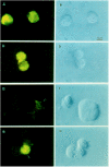
CXCR4 expression is reduced in Jurkat cells after induction of HIV-1 Env expression. After 4 days induction of HIV-1 Env proteins, non-induced and induced cells were fixed and labeled with a mouse MAb to CXCR4 (12G5) and a secondary FITC-conjugated anti-mouse antibodies for observation with a fluorescence microscopy. Panels A and C: CXCR4 staining of non-induced Jurakt cells. Panel E and G: CXCR4 staining of induced Jurkat cells. The fluorescent cell in panel G is representative of a minor population of cells in the induced culture (<5%) with a CXCR4 surface distribution similar to uninduced cells. Panels B, D, F and H show phase contrast images of the same fields of cells shown in left panels.

References

    1. Murphy PM. Viral exploitation and subversion of the immune system through chemokine mimicry. Nat Immunol. 2001;2:116–122. doi: 10.1038/84214. - DOI - PubMed
    1. Baggiolini M, Dewald B, Moser B. Human chemokines: an update. Annu Rev Immunol. 1997;15:675–705. doi: 10.1146/annurev.immunol.15.1.675. - DOI - PubMed
    1. Allen SJ, Crown SE, Handel TM. Chemokine: receptor structure, interactions, and antagonism. Annu Rev Immunol. 2007;25:787–820. doi: 10.1146/annurev.immunol.24.021605.090529. - DOI - PubMed
    1. Bacon K, Baggiolini M, Broxmeyer H, Horuk R, Lindley I, Mantovani A, Maysushima K, Murphy P, Nomiyama H, Oppenheim J, Rot A, Schall T, Tsang M, Thorpe R, Van Damme J, Wadhwa M, Yoshie O, Zlotnik A, Zoon K. Chemokine/chemokine receptor nomenclature. J Interferon Cytokine Res. 2002;22:1067–1068. doi: 10.1089/107999002760624305. - DOI - PubMed
    1. Feng Y, Broder CC, Kennedy PE, Berger EA. HIV-1 entry cofactor: functional cDNA cloning of a seven-transmembrane, G protein-coupled receptor [see comments] Science. 1996;272:872–877. doi: 10.1126/science.272.5263.872. - DOI - PubMed

Publication types

Substances