

Review
doi: 10.1016/j.drudis.2007.10.003.
Epub 2007 Nov 26.
Microfluidics for drug discovery and development: from target selection to product lifecycle management
Affiliations
- PMID: 18190858
- PMCID: PMC2700821
- DOI: 10.1016/j.drudis.2007.10.003
Item in Clipboard
Review
Microfluidics for drug discovery and development: from target selection to product lifecycle management
Drug Discov Today.
2008 Jan.
Abstract
Microfluidic technologies' ability to miniaturize assays and increase experimental throughput have generated significant interest in the drug discovery and development domain. These characteristics make microfluidic systems a potentially valuable tool for many drug discovery and development applications. Here, we review the recent advances of microfluidic devices for drug discovery and development and highlight their applications in different stages of the process, including target selection, lead identification, preclinical tests, clinical trials, chemical synthesis, formulations studies and product management.
Figures
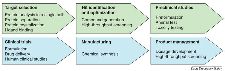
Drug discovery involves target selection, lead identification, optimization and preclinical studies. Drug development includes clinical trials, manufacturing, and product management process.
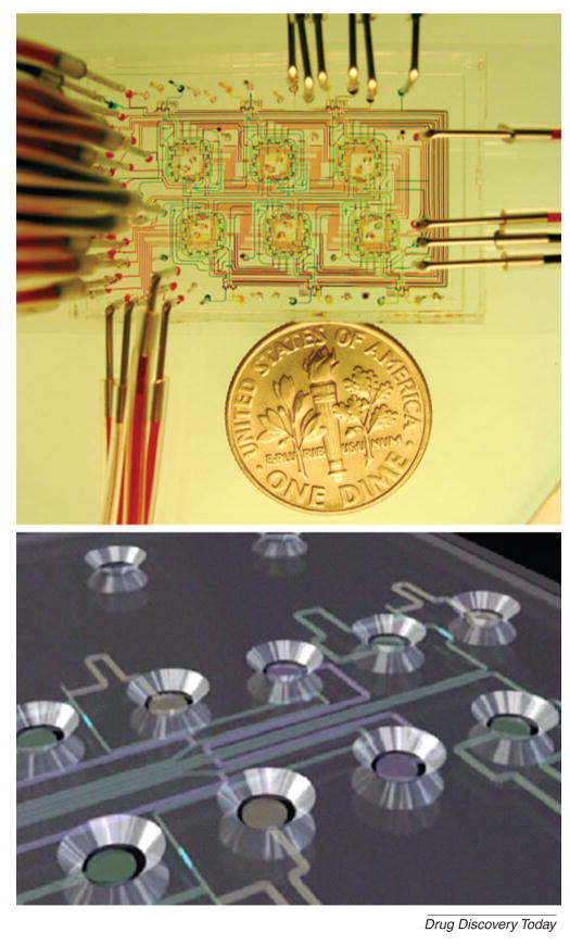
(A) A microfluidic chemostat device containing high-density on-chip valves used to study microbial growth. (Reprinted by permission from Macmillan Publishers Ltd: [Nature] (Whitesides, 442: 368–373) copyright 2006)[5]. (B) Schematic of Caliper LabChip device in which microchannels connect with individual circular microwells to screen drug candidates (Reprinted by permission from Macmillan Publishers Ltd: [Nature] (Smith et al., 446: 219–222) copyright 2007)[6].
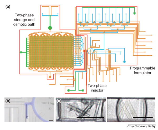
(A) Schematic of microfluidic screening platform with many addressable channels. (B) An aqueous droplet was introduced (blue) into an immiscible carrier fluid using a peristaltic pump. (C) Protein crystals were formed in a microfluidic channel. Crystals were generated from glucose isomerase (left) and catalase (right). Scale bar is 100 μm. (Reprinted with permission from Lau et al. Copyright 2006 American Chemical Society)[29].
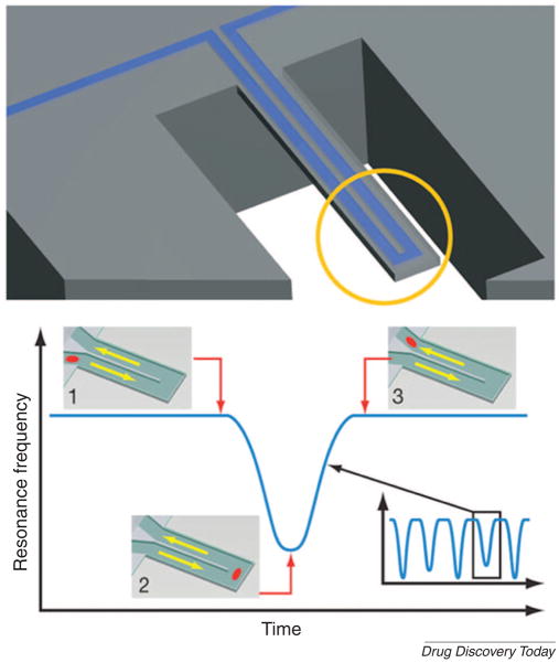
(A) Schematic design of a microfluidic cantilever that can be used to determine mass changes. (B) Particles flow through without binding to the surface. Mass changes of particles were quantified by measuring resonance frequency. (Reprinted by permission from Macmillan Publishers Ltd: [Nature] (Burg et al., 446: 1066–1069) copyright 2007)[38].
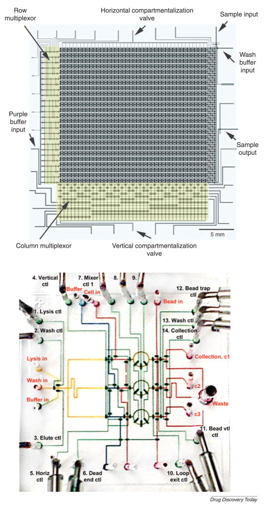
(A) A high-density microfluidic device integrated with microfluidic multiplexors. Valves are formed at the intersection of control channels with fluidic channels. (Reprinted with permission from Thorsen et al. Copyright 2002 Science)[53]. (B) DNA purification chip. This device is used for multiple parallel processes of DNA recovery from living bacterial cells. (Reprinted by permission from Macmillan Publishers Ltd: [Nature Biotechnology] (Hong et al., 22: 435–439) copyright 2004)[55].
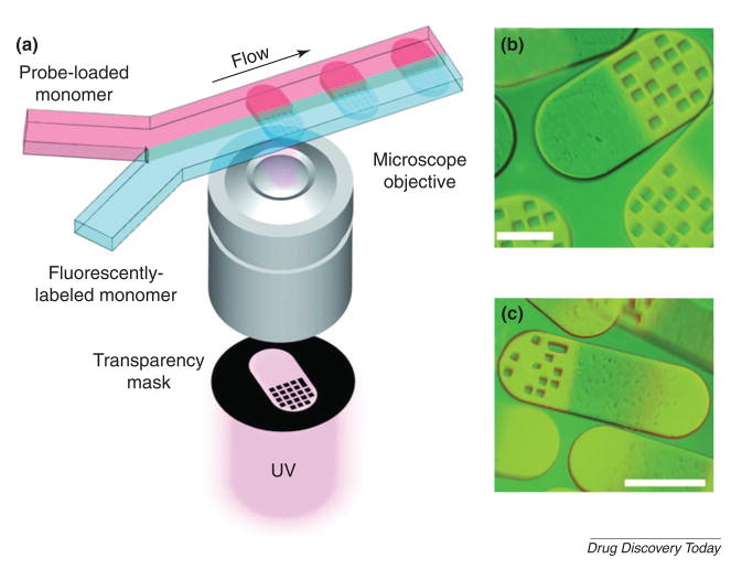
(A) Schematic diagram of particle synthesis through photo-polymerization across laminar streams. Fluorescence images of single-probe (B) and probe-gradient (C). Scale bar is 100 μm. (Reprinted with permission from Pregibon et al. Copyright 2007 Science)[56].
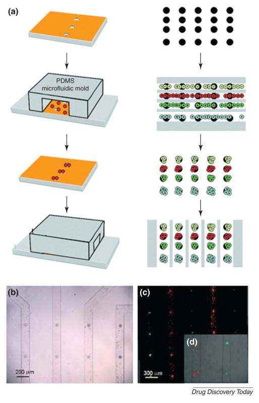
(A) Schematic drawing of reversibly sealed microfluidic device that can be aligned on an array of microwells. (B) Embryonic stem cells could be docked within the microchannels. (C) fluorescent image of cells labeled with membrane dyes CFSE (green) and SYTO (red). (D) A cell-free solution is flowed through the channels to remove non-adhered cells. (Khademhosseini et al. Lab Chip, 5, 1380–1386) -Reproduced by permission of The Royal Society of Chemistry[60].
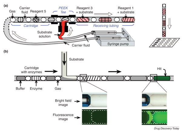
(A) The experimental setup using preloaded cartridges of nanoliter plugs for screening reagents. After incubation, the plugs are deposited onto a plate for analyzing and screening reagents. (B) Schematic drawing of a fluorescence assay using microfluidic cartridges with plugs with enzymes. (Reprinted with permission from Chen et al. Copyright 2006 Elsevier)[62].
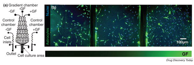
(A) Schematic of the microfluidic device integrated with control and gradient chambers. (B) Differentiation of human NSCs exposed to gradients of growth factor mixtures in the microfluidic device. Fluorescence images show human NSCs stained with an astrocytes marker (green) and nuclear stain (blue). [Chung et al. Lab Chip, 5, 401–406] - Reproduced by permission of The Royal Society of Chemistry[66].
References
-
- Editorial. Five years on…and four challenges for the pharmaceutical industry. Nat Rev Drug Discov. 2007;6:3. - PubMed
-
- Editorial. Same old story? Nat Rev Drug Discov. 2007;6:97. - PubMed
-
- Owens J. 2006 drug approvals: finding the niche. Nat Rev Drug Discov. 2007;6:99–101. - PubMed
-
- PhRMA. Drug discovery and development: Understanding the R&D process. 2007. http://www.innovation.org/drug_discovery/objects/pdf/RD_Brochure.pdf.
-
- Whitesides GM. The origins and the future of microfluidics. Nature. 2006;442:368–373. - PubMed
Publication types
MeSH terms
Grants and funding
LinkOut - more resources
Full Text Sources
Other Literature Sources
