Review
. 2008 May;53(5):596-612.
doi: 10.1016/j.yhbeh.2007.11.008. Epub 2007 Nov 28.

The spinal nucleus of the bulbocavernosus: firsts in androgen-dependent neural sex differences

Affiliations
Review

The spinal nucleus of the bulbocavernosus: firsts in androgen-dependent neural sex differences

Dale R Sengelaub et al. Horm Behav. 2008 May.

Abstract

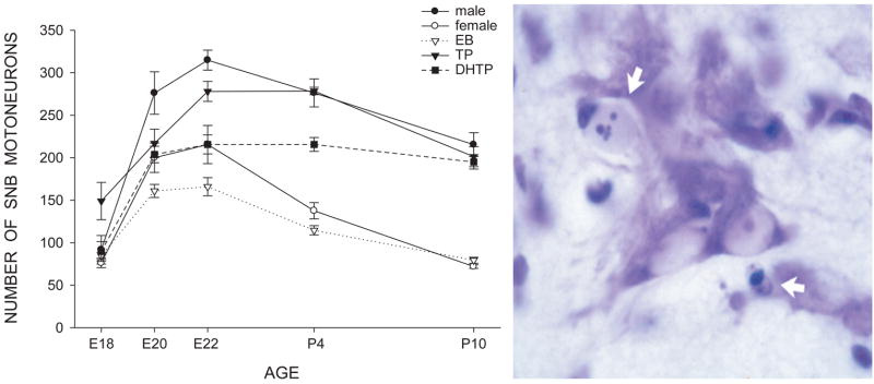
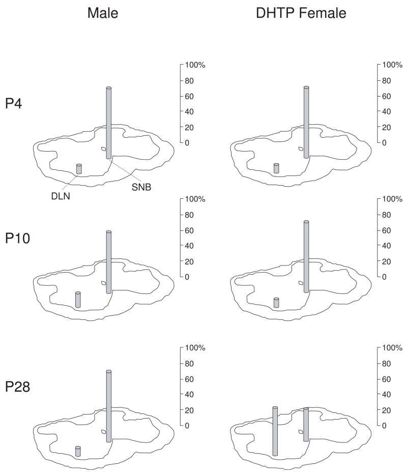
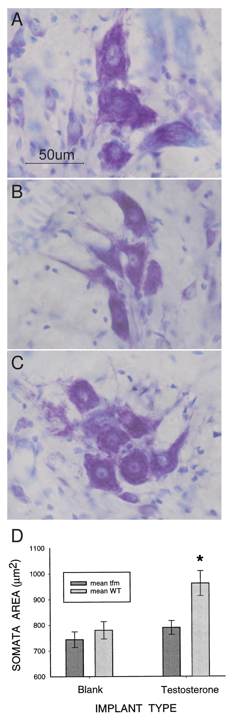
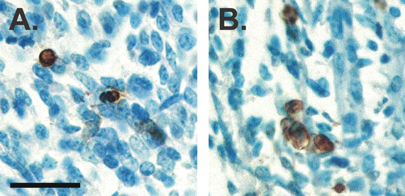
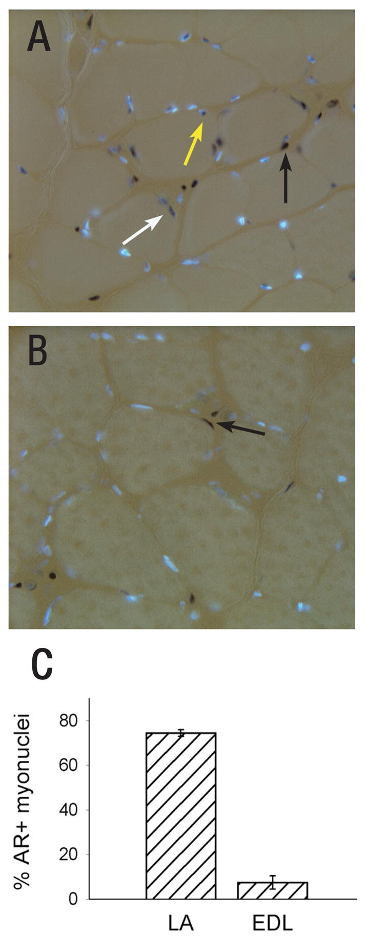
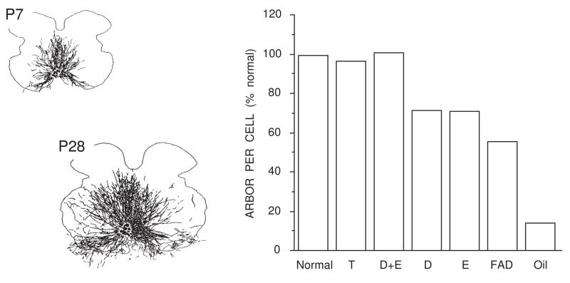
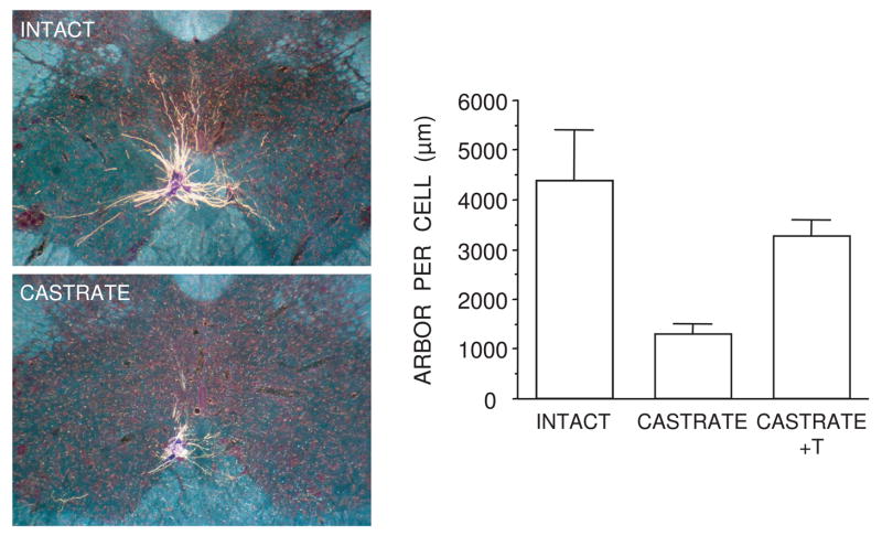
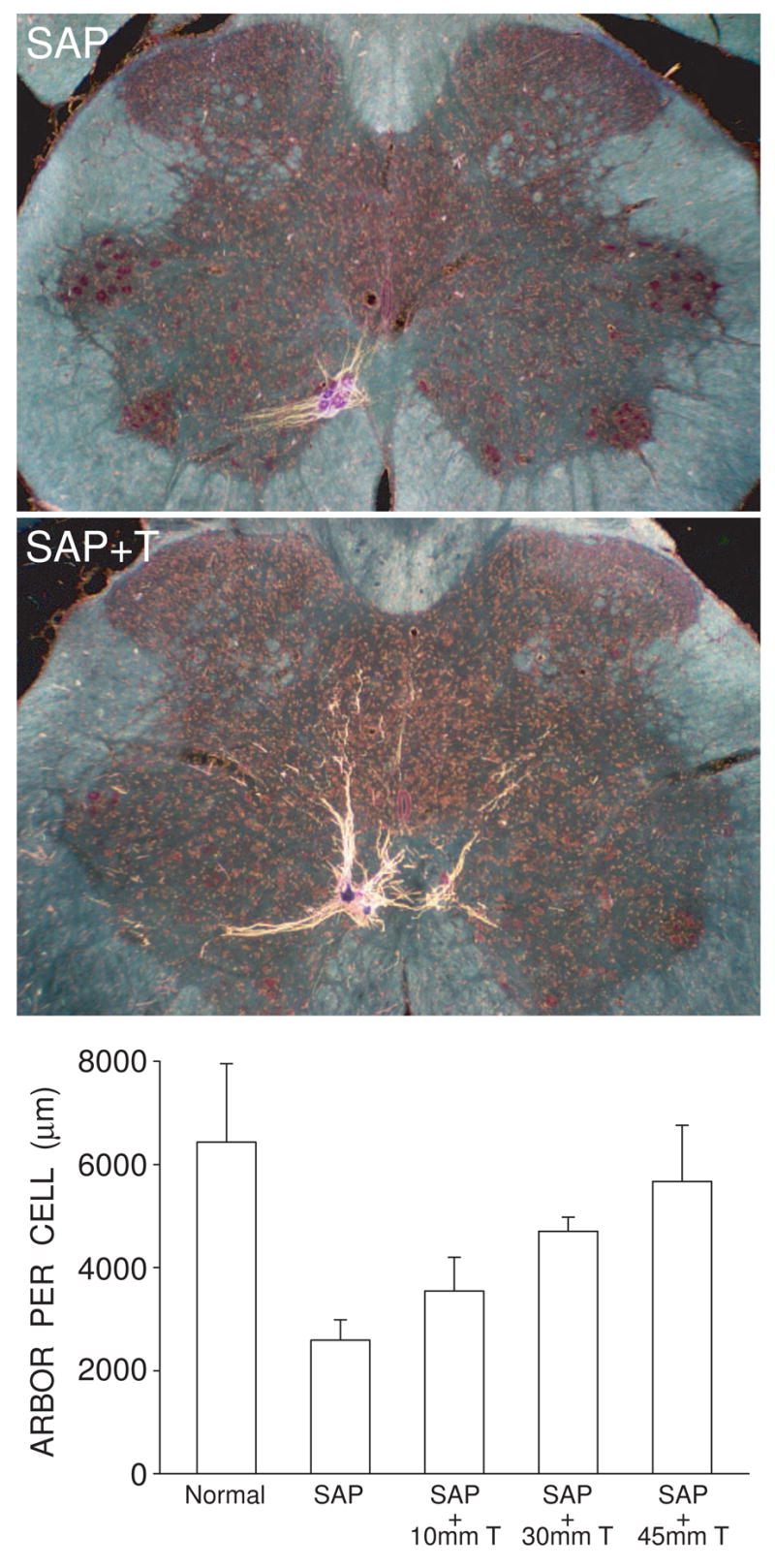
Cell number in the spinal nucleus of the bulbocavernosus (SNB) of rats was the first neural sex difference shown to differentiate under the control of androgens, acting via classical intracellular androgen receptors. SNB motoneurons reside in the lumbar spinal cord and innervate striated muscles involved in copulation, including the bulbocavernosus (BC) and levator ani (LA). SNB cells are much larger and more numerous in males than in females, and the BC/LA target muscles are reduced or absent in females. The relative simplicity of this neuromuscular system has allowed for considerable progress in pinpointing sites of hormone action, and identifying the cellular bases for androgenic effects. It is now clear that androgens act at virtually every level of the SNB system, in development and throughout adult life. In this review we focus on effects of androgens on developmental cell death of SNB motoneurons and BC/LA muscles; the establishment and maintenance of SNB motoneuron soma size and dendritic length; BC/LA muscle morphology and physiology; and behaviors controlled by the SNB system. We also describe new data on neurotherapeutic effects of androgens on SNB motoneurons after injury in adulthood.


Figures

Figure 1
Figure 1
Development of SNB motoneuron number is regulated through an androgen-mediated normally occurring cell death. (Left) Counts of motoneurons in the SNB from embryonic day (E)18 through postnatal day (P)10 for normal males and females, as well as females treated both pre- and postnatally with either estradiol benzoate- (EB), testosterone propionate- (TP), and dihydrotestosterone propionate- (DHTP). Females treated with TP or DHTP have a masculine number of SNB cells on P10, whereas females treated with EB do not differ from controls. Points represent means ± SEM. (Right) Photomicrograph of a transverse, cresylecht violet-stained section through the SNB at P2 showing both normal motoneurons and degenerating cells (arrows). (Compiled from data originally published in Nordeen et al., 1985, Goldstein and Sengelaub, 1990, .)
Figure 2
Figure 2
Relative proportions of motoneurons in the SNB and DLN retrogradely labeled via the BC muscle at postnatal (P) days 4, 10, and 28 in both normal males (left) and females treated prenatally with dihydrotestosterone propionate (DHTP; right). Motoneurons in normal males, and females cross-fostered from pregnant dams treated on prenatal days 17–22 with DHTP (4 mg/day) were labeled following unilateral injections of horseradish peroxidase conjugated to the cholera toxin B subunit (BHRP; 0.05 μL, 0.2%) into the BC muscle on embryonic (E) day 22, and days P4, P10, and P28 (n=5–9 per group); post-injection survival times varied by age (6 hours at E22, 24 hours at P4 and P10, 48 hours at P28). Counts of labeled motoneurons in the DLN and SNB were made under bright-field illumination at 500X and corrected by the method of Konigsmark (1970). On P4 and P10, the majority of the retrogradely labeled motoneurons are found in the SNB in both males and DHTP-treated females. By P28, however, the number of motoneurons in the DLN projecting to the BC muscle in DHTP-treated females has substantially increased. (Data from Kalkbrenner and Sengelaub, 1992.)
Figure 3
Figure 3
Digital micrographs of thionin-stained SNB somata in a normal adult male (A), a castrate (B), and a castrate treated with testosterone (C), demonstrating the androgen sensitivity of soma size in adulthood. (D) Testosterone increases SNB soma size only in cells expressing the wild-type (WT) androgen receptor (AR). Adult female carriers of the Tfm mutation were implanted with blank capsules or capsules filled with testosterone. SNB soma size was assessed 4–6 weeks later. Some SNB motoneurons in these females express the WT AR, whereas others express the non-functional, Tfm AR. Only SNB motoneurons expressing the WT AR responded to testosterone with an increase in soma size. (Panels A–C, D.R. Sengelaub; Panel D reprinted from Watson et al., (2001) with permission from the Society for Neuroscience.)
Figure 4
Figure 4
Photomicrographs of sections through the LA muscle of a male (A) and female (B) mouse on embryonic day 18. Tissue was stained with TUNEL to label dying cells. The density of TUNEL-positive cells (brown) is about two-fold higher in females than in males. Scale bar = 25 μm. (Panels A and B, N. Forger.)
Figure 5
Figure 5
Androgen receptor (AR) expression is unusually high in myofibers of the LA. In the photomicrographs shown in (A) and (B), DAPI (blue) labels AR negative nuclei and black peroxidase labels AR+ nuclei. A fluorescent basal lamina stain (not shown) was used to discriminate between AR in muscle fiber nuclei (white and yellow arrows) or in fibroblasts, which are found in the interstitial space between fibers (black arrows). (A) Cross section through the LA reveals many AR+ myonuclei; (B) Cross section through the extensor digitorum longus (EDL) limb muscle shows few AR+ myonuclei. (C) The percentage of AR+ muscle fibers in the LA and EDL. (Reprinted from Monks et al. (2004) with permission from John Wiley and Sons.)
Figure 6
Figure 6
SNB dendritic growth requires both androgens and estrogens for normal masculine development. (Left) Computer-generated composites of retrogradely-labeled somata and processes drawn at 320 μm intervals through the entire rostrocaudal extent of the SNB of normal males at P7 (top) and P28 (bottom), illustrating the exuberant growth of SNB dendrites. (Right) SNB dendritic lengths at P28 in intact males, postnatally castrated males treated with testosterone propionate (T), dihydrotestosterone propionate combined with estradiol benzoate (D+E), D or E alone, gonadally intact males treated with fadrozole (FAD), and oil-treated castrates. Data are expressed as a percentage of dendritic length in intact males. (Compiled from data originally published in Goldstein et al., 1990, Goldstein and Sengelaub, 1994, Burke et al., 1997, .)
Figure 7
Figure 7
SNB dendritic maintenance in adulthood requires androgens. (Left) Darkfield digital micrographs of transverse sections through the lumbar spinal cord of an adult normal male (top) and a castrated male (bottom) after injection of horseradish peroxidase conjugated to the cholera toxin B subunit (BHRP), into the left BC muscle. (Right) SNB dendritic lengths expressed as length of arbor per labeled motoneuron for gonadally intact males, castrated males, and castrates treated with testosterone (T). Bar heights represent means ± SEM. (Data after Kurz et al., 1986.)
Figure 8
Figure 8
Testosterone treatment is neurotherapeutic in SNB motoneurons, attenuating induced dendritic atrophy resulting from the death of nearby motoneurons. (Left) Darkfield digital micrographs of transverse sections through the lumbar spinal cords of males whose SNB motoneurons have been partially depleted with the toxin saporin, and then left untreated (SAP) or given supplemental testosterone (SAP + T), after BHRP injection into the left BC muscle. (Right) SNB dendritic lengths expressed as length of arbor per labeled motoneuron for normal males and saporin-treated males with or without supplemental testosterone. Partial motoneuron depletion reduces the dendritic arbor of remaining motoneurons. However, in rats whose androgen levels had been supplemented with varying levels of T, this atrophy is attenuated in a dose-dependent manner. Bar heights represent means ± SEM. (Data from Coons et al., 2007.)

Similar articles

Cited by

References

    1. Araki I, Harada Y, Kuna M. Target-dependent hormonal control of neuron size in the rat spinal nucleus of the bulbocavernosus. J Neurosci. 1991;11:3025–3033. - PMC - PubMed
    1. Balice-Gordon RJ, Breedlove SM, Bernstein S, Lichtman JW. Neuromuscular junctions shrink and expand as muscle fiber size is manipulated: in vivo observations in the androgen-sensitive bulbocavernosus muscle of mice. J Neurosci. 1990;10:2660–2671. - PMC - PubMed
    1. Bengston L, Lopez V, Watamura S, Forger NG. Short- and long-term effects of ciliary neurotrophic factor on androgen-sensitive motoneurons in the lumbar spinal cord. J Neurobiol. 1996;31:263–273. - PubMed
    1. Bleisch WV, Harrelson A. Androgens modulate endplate size and ACh receptor density at synapses in rat levator ani muscle. J Neurobiol. 1989;20:189–202. - PubMed
    1. Bleisch WV, Harrelson AL, Luine VN. Testosterone increases acetylcholine receptor number in the “levator ani” muscle of the rat. J Neurobiol. 1982;13:153–161. - PubMed

Publication types