. 2008 May;44(5):426-34.
doi: 10.1016/j.micpath.2007.11.006. Epub 2007 Nov 28.

Staphylococcus aureus lipoproteins trigger human corneal epithelial innate response through toll-like receptor-2

Affiliations

Staphylococcus aureus lipoproteins trigger human corneal epithelial innate response through toll-like receptor-2

Qiong Li et al. Microb Pathog. 2008 May.

Abstract

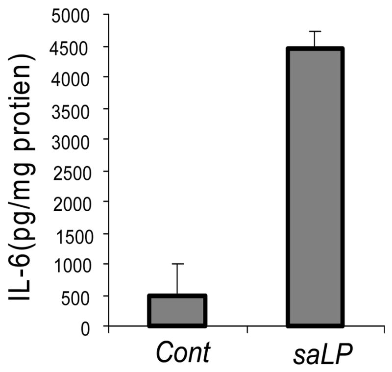
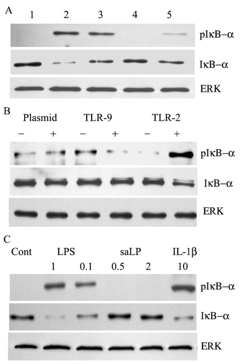
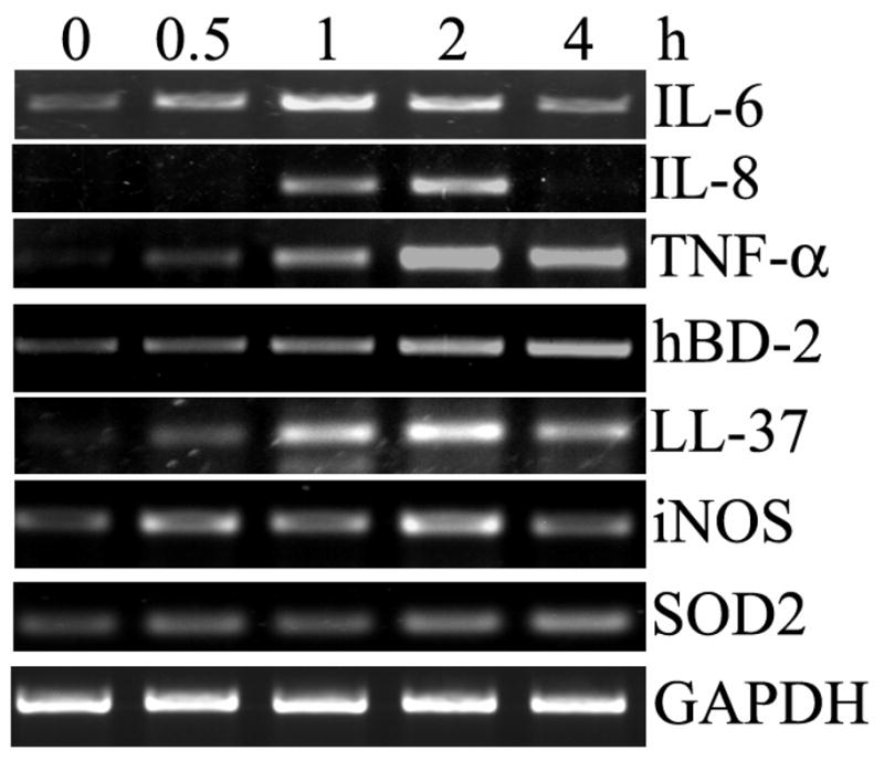
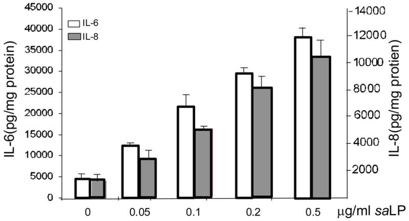
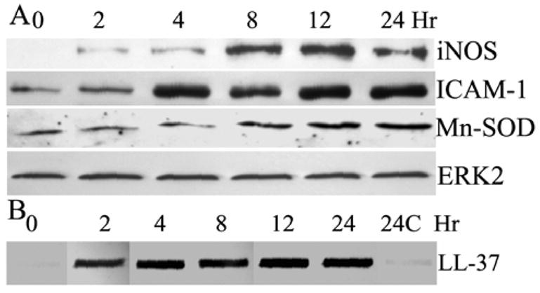
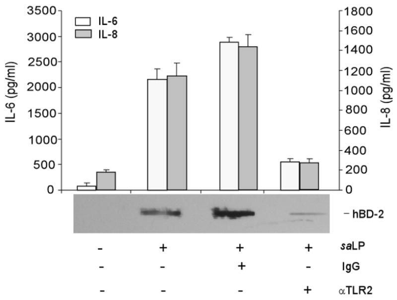
Bacterial lipoproteins (LP) are a family of cell wall components found in a wide variety of bacteria. In this study, we characterized the response of HUCL, a telomerase-immortalized human corneal epithelial cell (HCEC) line, to LP isolated from Staphylococcus (S) aureus. S. aureus LP (saLP) prepared by Triton X-114 extraction stimulated the activation of NF-kappaB, JNK, and P38 signaling pathways in HUCL cells. The extracts failed to stimulate NF-kappaB activation in HUCL cells after lipoprotein lipase treatment and in cell lines expressing TLR4 or TLR9, but not TLR2, indicating lipoprotein nature of the extracts. saLP induced the up-regulation of a variety of inflammatory cytokines and chemokines (IL-6, IL-8, ICAM-1), antimicrobial molecules (hBD-2, LL-37, and iNOS), and homeostasis genes (Mn-SOD) at both the mRNA level and protein level. Similar inflammatory response to saLP was also observed in primarily cultured HCECs using the production of IL-6 as readout. Moreover, TLR2 neutralizing antibody blocked the saLP-induced secretion of IL-6, IL-8 and hBD2 in HUCL cells. Our findings suggest that saLP activates TLR2 and triggers innate immune response in the cornea to S. aureus infection via production of proinflammatory cytokines and defense molecules.


Figures

Figure 1
Figure 1. Triton X-114 extract-induced IL-6 secretion in primary HCECs
Primary HCECs were stimulated with (saLP) or without (Cont) Triton X-114 extracts of S. aureus (0.5 μg/ml protein) for 10 h. The supernatants were collected and IL-6 secretion was measured by ELISA. The amount of IL-6 in the culture media was normalized with the protein concentration of cell lysates. Results shown are representative of two independent experiments, *P < 0.001, N=3.
Figure 2
Figure 2. saLP-induced activation of NF-κB as well P38 and JNK pathways
(A) HUCL cells were stimulated with different concentrations of saLP for 1h and total protein was extracted and subjected to SDS-PAGE, followed by phospho-IκB-α (pIκB-α) and IκB-α immunoblotting. (B) HUCL cells were stimulated with 0.5 μg/ml saLP for indicated time (hours) and cells were lysed for Western blot analysis with antibodies against phospho-IκB-α (pIκB-α), IκB-α, phospho-p38 (pP38), phospho-JNK (pJNK). Total ERK was used as protein loading control. Results shown are representative of three independent experiments.
Figure 3
Figure 3. Lipoprotein lipase sensitive and TLR-dependent activation of NF-κB induced by saLP
(A) HUCL cells were challenged with saLP (lane 2), saLP digested with (lane 5) or without (lane lane 3) lipoprotein lipase (8500unit/ml, 37°C, 6 h); or lipoprotein lipase (lane 4), along with HUCL cells without treatment as the control (Lane1). After 1 h incubation, total protein was extracted for Western blotting to detect phospho-IκB-α and IκB-α. (B) Stable HEK cell lines transfected with empty (plasmid), TLR2, or TLR9 plasmids were stimulated with (+) or without (−) 0.5 μg/ml saLP. After 1 h incubation, cells were lysed and subjected to phospho-IκB-α and IκB-α. (C) Stable Hela cell line expressing TLR4, CD14 and MD2 were stimulated with LPS (1 and 0.1 μg/ml), or saLP (0.5 or 2 μg/ml), with the controls including unstimulated (Cont) or 10 ng/ml IL-1β. After 1 h incubation, cells were lyased and subjected to Western blotting to detect phospho-IκB-α and IκB-α. Total ERK was used as protein loading control. The results are representative for two independent experiments.
Figure 4
Figure 4. saLP-elicited expression of proinflammatory cytokine/chemokine, antimicrobial molecules, and Mn-SOD in HUCL cells
HUCL were incubated with 0.5 μg/ml saLP for the indicated times (hours) and the total cellular RNA was extracted at the end of incubation and subjected to RT-PCR analysis with primers amplifying human IL-6, IL-8, TNF-α, hBD-2, LL-37 iNOS, Mn-SOD with GAPDH as the internal control. The results are representative of three independent experiments.
Figure 5
Figure 5. saLP concentration dependent induction of IL-6 and IL-8 production in HUCL cells
HUCL cells were challenged with increasing concentrations of saLP for 8 h along with HUCL cells without stimulation as the control. Accumulated levels of IL-6 and IL-8 in cell culture supernatant were measured by ELISA. Statistical analysis was performed using one way ANOVA (N=3; P=0.0015 for IL-6 and 0.002 for IL-8). Data are representative of two independent experiments.
Figure 6
Figure 6. saLP-induced protein expression of iNOS, ICAM-1, Mn-SOD and accumulation of LL-37 in HUCL cells
HUCL cells were stimulated with 0.5 μg/ml saLP for the indicated time (hours). At the end of incubation, the culture media were collected and cells were lysed. The cell lysates were subjected to Western blotting analysis with antibodies against ICAM-1, iNOS and Mn-SOD (A). The collected culture media were assayed for LL-37 production by slot-blot (B) with both media collected from untreated cells at beginning (0 h) and after 24 h of culture (24C) as the controls. The results are representative for two independent experiments.
Figure 7
Figure 7. saLP induced IL-6, IL-8, and hBD-2 secretion in HUCL cells were TLR2 dependent
HUCL cells were pre-incubated with 20 μg/ml anti-TLR2 or mouse IgGα as the control antibody for 1 h. Antibody-pretreated cells were incubated with 0.5 μg/ml saLP for 8h. Culture supernatant was analyzed for IL-6 and IL-8 production by ELISA or for hBD2 accumulation by slot-blot. Statistical analysis of IL-6 and IL-8 secretion was performed using one way ANOVA (N=3, P=0.0048 for IL-6 and 0.0056 for IL-8). Data shown are representative of two independent experiments.

References

    1. Sun X, Deng S, Li R, Wang Z, Luo S, Jin X, Zhang W. Distribution and shifting trends of bacterial keratitis in north China (1989–98) Br J Ophthalmol. 2004;88:165–166. - PMC - PubMed
    1. Alexandrakis G, Alfonso EC, Miller D. Shifting trends in bacterial keratitis in south Florida and emerging resistance to fluoroquinolones. Ophthalmology. 2000;107:1497–1502. - PubMed
    1. Jett BD, Gilmore MS. Host-parasite interactions in Staphylococcus aureus keratitis. DNA Cell Biol. 2002;21:397–404. - PubMed
    1. Cohen E, Laibson P, Arentsen J, Clemons C. Corneal ulcers associated with cosmetic extended wear soft contact lenses. Ophthalmology. 1987;94:109–114. - PubMed
    1. Kurpakus-Wheater M, Kernacki KA, Hazlett LD. Maintaining corneal integrity how the “window” stays clear. Prog Histochem Cytochem. 2001;36:185–259. - PubMed

Publication types

MeSH terms