. 2008 May 2;283(18):12212-26.
doi: 10.1074/jbc.M709355200. Epub 2008 Jan 10.

Mutational analysis of Deinococcus radiodurans bacteriophytochrome reveals key amino acids necessary for the photochromicity and proton exchange cycle of phytochromes

Affiliations

Mutational analysis of Deinococcus radiodurans bacteriophytochrome reveals key amino acids necessary for the photochromicity and proton exchange cycle of phytochromes

Jeremiah R Wagner et al. J Biol Chem. .

Abstract

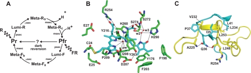
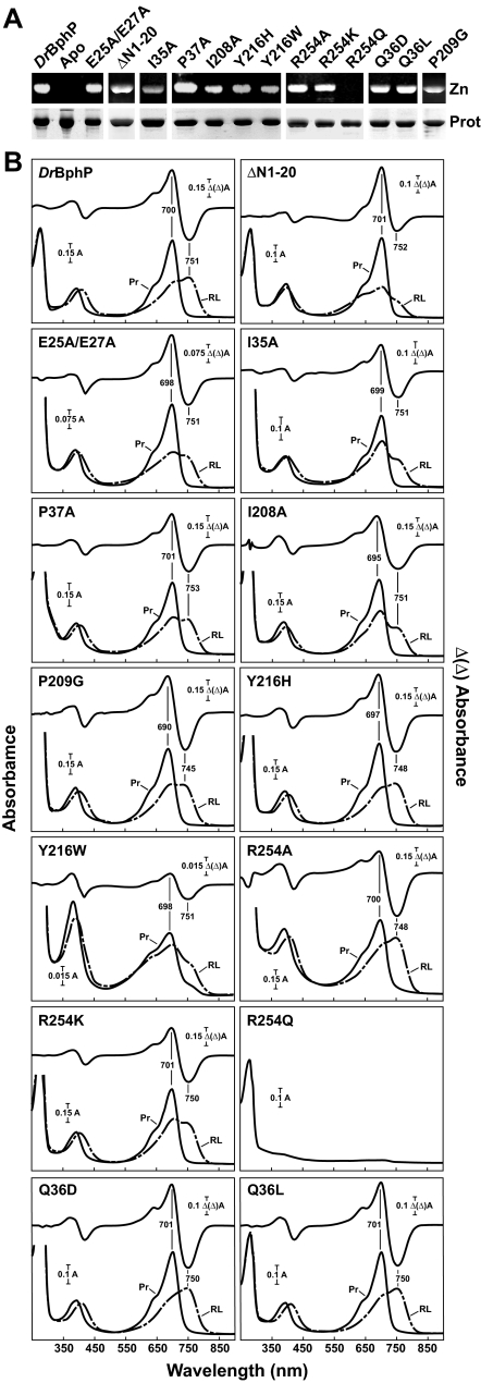
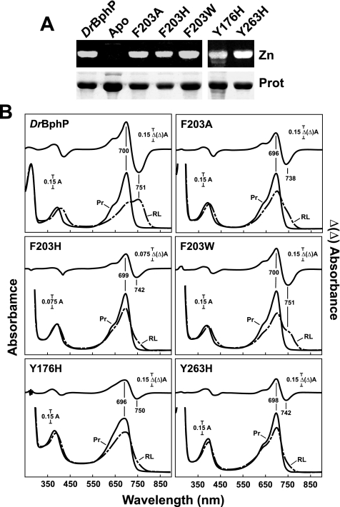
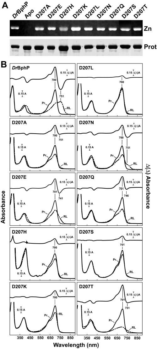
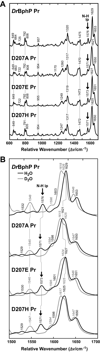
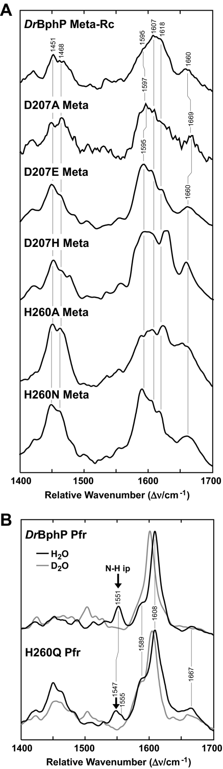
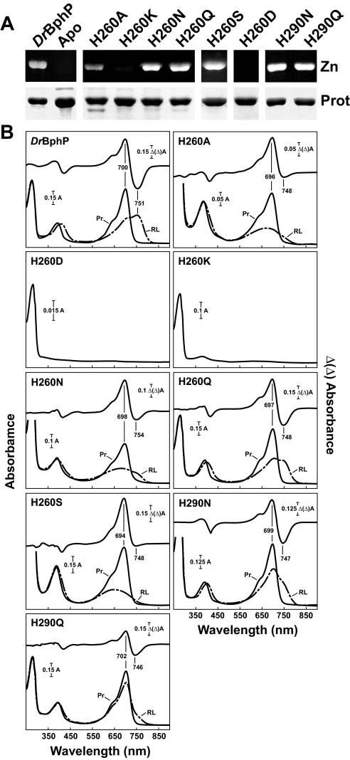
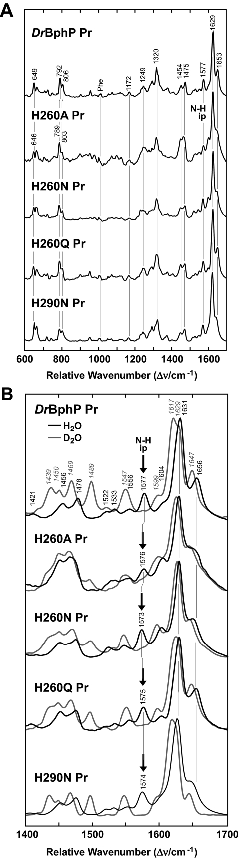
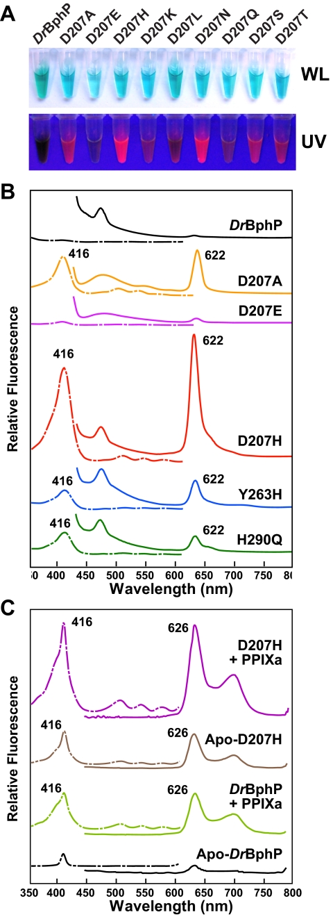
The ability of phytochromes (Phy) to act as photointerconvertible light switches in plants and microorganisms depends on key interactions between the bilin chromophore and the apoprotein that promote bilin attachment and photointerconversion between the spectrally distinct red light-absorbing Pr conformer and far red light-absorbing Pfr conformer. Using structurally guided site-directed mutagenesis combined with several spectroscopic methods, we examined the roles of conserved amino acids within the bilin-binding domain of Deinococcus radiodurans bacteriophytochrome with respect to chromophore ligation and Pr/Pfr photoconversion. Incorporation of biliverdin IXalpha (BV), its structure in the Pr state, and its ability to photoisomerize to the first photocycle intermediate are insensitive to most single mutations, implying that these properties are robust with respect to small structural/electrostatic alterations in the binding pocket. In contrast, photoconversion to Pfr is highly sensitive to the chromophore environment. Many of the variants form spectrally bleached Meta-type intermediates in red light that do not relax to Pfr. Particularly important are Asp-207 and His-260, which are invariant within the Phy superfamily and participate in a unique hydrogen bond matrix involving the A, B, and C pyrrole ring nitrogens of BV and their associated pyrrole water. Resonance Raman spectroscopy demonstrates that substitutions of these residues disrupt the Pr to Pfr protonation cycle of BV with the chromophore locked in a deprotonated Meta-R(c)-like photoconversion intermediate after red light irradiation. Collectively, the data show that a number of contacts contribute to the unique photochromicity of Phy-type photoreceptors. These include residues that fix the bilin in the pocket, coordinate the pyrrole water, and possibly promote the proton exchange cycle during photoconversion.


Figures

FIGURE 1.
FIGURE 1.
Schematic presentation of the photocycle of Phys (A) and three-dimensional relationships of key amino acids within the CBD structure of DrBphP (B and C). B, region surrounding the BV chromophore. BV is colored cyan. Sulfur, oxygen, and nitrogen atoms are colored yellow, red, and purple, respectively. Pyrrole water (pw), water2 (w2), and water3 (w3) are indicated. C, region surrounding the knot interface. The PAS and GAF polypeptide chains are colored cyan and yellow, respectively. Dashed lines highlight key noncovalent interactions. Three-dimensional structures are from Refs. , .
FIGURE 2.
FIGURE 2.
Assembly and absorption spectra of DrBphP mutants potentially affecting bilin binding and the knot interface. The recombinant full-length and ΔN1–20 truncated apoproteins were incubated with BV and purified by nickel chelate chromatography. A, samples were subjected to SDS-PAGE and either assayed for the bound bilin by zinc-induced fluorescence (Zn) or stained for protein with Coomassie Blue (Prot). Apo, apoprotein prior to BV incubation. B, UV-visible absorption spectra as Pr (Pr, black lines) or following saturating red light irradiation (RL, dashed lines) and red-minus-far red light difference spectra (upper graph) of the apoproteins incubated with BV. Difference spectra maxima and minima are indicated.
FIGURE 3.
FIGURE 3.
Assembly and absorption spectra of amino acid substitutions in DrBphP affecting bulky hydrophobic residues near the D ring of BV. The recombinant full-length apoproteins were incubated with BV and purified by nickel chelate chromatography. A, samples were subjected to SDS-PAGE and either assayed for the bound bilin by zinc-induced fluorescence (Zn) or stained for protein with Coomassie Blue (Prot). Apo, apoprotein prior to BV incubation. B, UV-visible absorption spectra as Pr (Pr, black lines) or following saturating red light irradiation (RL, dashed lines) and red-minus-far red light difference spectra (upper graph) of the apoproteins incubated with BV. Difference spectra maxima and minima are indicated.
FIGURE 4.
FIGURE 4.
Assembly and absorption spectra of amino acid substitutions in DrBphP affecting Asp-207. The recombinant full-length apoproteins were incubated with BV and purified by nickel chelate chromatography. A, samples were subjected to SDS-PAGE and either assayed for the bound bilin by zinc-induced fluorescence (Zn) or stained for protein with Coomassie Blue (Prot). Apo, apoprotein prior to BV incubation. B, UV-visible absorption spectra as Pr (Pr, black lines) or following saturating red light irradiation (RL, dashed lines) and redminus-far red light difference spectra (upper graph) of the apoproteins incubated with BV. Difference spectra maxima and minima are indicated.
FIGURE 5.
FIGURE 5.
RR spectra of the Pr state of DrBphP and the D207A, D207E, and D207H variants. A, overview RR spectra (600–1700 cm–1) measured from the samples dissolved in H2O. B, expanded view of the spectra in A for the marker band region (1500–1700 cm–1) from samples dissolved in H2O(black lines) and D2O(gray lines). The positions of N-H ip band (arrows) and other diagnostic RR maxima are indicated.
FIGURE 6.
FIGURE 6.
RR spectra of the photoconversion products of DrBphP and variants affecting Asp-207 and His-260. A, RR spectra of wild-type DrBphP in the Meta-Rc state trapped at –30 °C during red light irradiation as compared with the meta states of D207A, D207E, D207H, H260A, and H260N variants formed at room temperature during red light irradiation. B, RR spectra of Pfr state of wild-type DrBphP and the H260Q mutant measured from samples either dissolved in H2O(black lines) and D2O(gray lines) and then irradiated at room temperature with red light. Contributions from the residual Pr state were subtracted from each spectrum. The positions of N-H ip band (arrows) and other diagnostic RR maxima are indicated.
FIGURE 7.
FIGURE 7.
Assembly and absorption spectra of amino acid substitutions in DrBphP affecting His-260 and His-290. The recombinant full-length apoproteins were incubated with BV and purified by nickel chelate chromatography. A, samples were subjected to SDS-PAGE and either assayed for the bound bilin by zinc-induced fluorescence (Zn) or stained for protein with Coomassie Blue (Prot). Apo, apoprotein prior to BV incubation. B, UV-visible absorption spectra as Pr (black lines) or following saturating red light irradiation (dashed lines) and red-minus-far red light difference spectra (upper graph) of the apoproteins incubated with BV. Difference spectra maxima and minima are indicated.
FIGURE 8.
FIGURE 8.
RR spectra of the Pr state of amino acid substitutions in DrBphP affecting His-260 and His-290. A, overview RR spectra (600–1700 cm–1) measured from the samples dissolved in H2O. B, expanded view of the spectra in A for the marker band region (1400–1700 cm–1) from samples dissolved in H2O(black lines) and D2O(gray lines). The positions of N-H ip band (arrows) and other diagnostic RR maxima are indicated.
Figure 9.
Figure 9.
Asp-207, His-290, and Tyr-263 mutants bind porphyrin and become red fluorescent. The UV-vis absorption spectra of each variant assembled with BV are shown in Figs. 3, 4, and 7. A, purified solutions of the DrBphP and the various Asp-207 variants in white light (WL) or upon irradiation with UV light. The concentration of each sample was adjusted to have an absorbance at ∼700 nm of 0.5 for the Pr form. B, fluorescence excitation (dashed lines) and emission spectra (solid lines) of the wild-type and mutant DrBphP after in vivo assembly with BV. Sample concetrations were adjusted to have an absorbance at 700 nm of 0.6 for the Pr form. C, fluorescence excitation (dashed lines) and emission spectra (solid lines) of purified wild-type DrBphP and the D207H mutant before (Apo) and after incubation with PPIXa in vitro. Sample concentrations were adjusted to have an absorbance at 280 nm of 2.

References

    1. Rockwell, N. C., Su, Y. S., and Lagarias, J. C. (2006) Annu. Rev. Plant Biol. 57 837–858 - PMC - PubMed
    1. Vierstra, R. D., and Karniol, B. (2005) in Handbook of Photosensory Receptors (Briggs, W. R., and Spudich, J. L., eds) pp. 171–196, Wiley-VCH Press, Weinheim, Germany
    1. Quail, P. H. (2002) Nat. Rev. Mol. Cell Biol. 3 85–93 - PubMed
    1. Yeh, K. C., and Lagarias, J. C. (1998) Proc. Natl. Acad. Sci. U. S. A. 95 13976–13981 - PMC - PubMed
    1. Karniol, B., Wagner, J. R., Walker, J. M., and Vierstra, R. D. (2005) Biochem. J. 392 103–116 - PMC - PubMed

Publication types