. 2008 Apr 15;94(8):3023-34.
doi: 10.1529/biophysj.107.118893. Epub 2008 Jan 11.

Alpha2delta1 dihydropyridine receptor subunit is a critical element for excitation-coupled calcium entry but not for formation of tetrads in skeletal myotubes

Affiliations

Alpha2delta1 dihydropyridine receptor subunit is a critical element for excitation-coupled calcium entry but not for formation of tetrads in skeletal myotubes

Marcin P Gach et al. Biophys J. .

Abstract

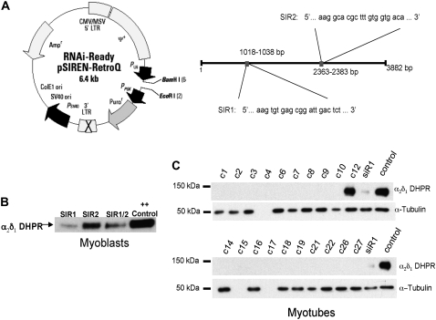
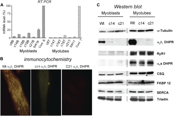
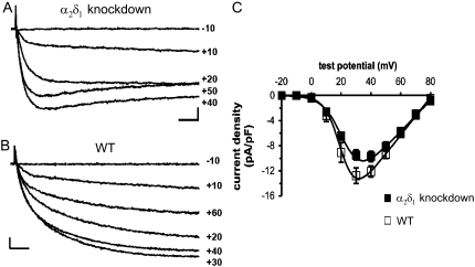
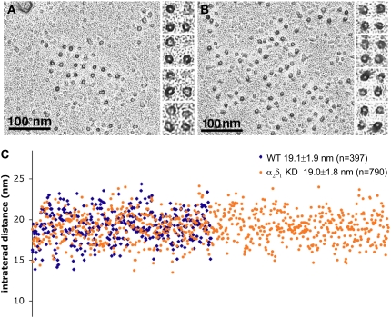
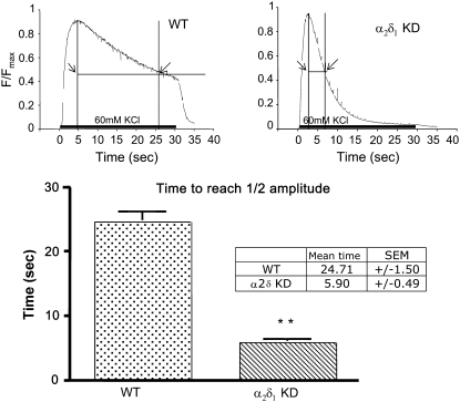
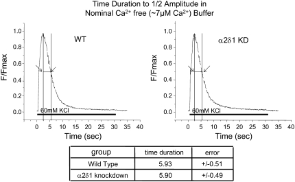
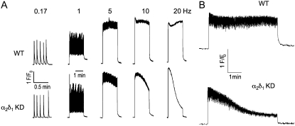
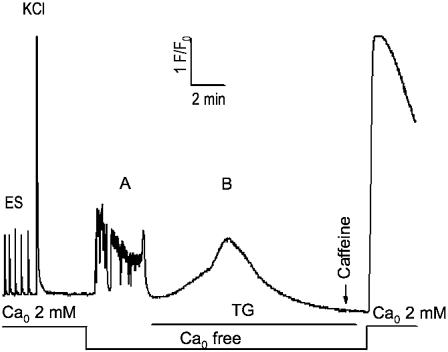
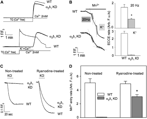
It has been shown that small interfering RNA (siRNA) partial knockdown of the alpha(2)delta(1) dihydropyridine receptor subunits cause a significant increase in the rate of activation of the L-type Ca(2+) current in myotubes but have little or no effect on skeletal excitation-contraction coupling. This study used permanent siRNA knockdown of alpha(2)delta(1) to address two important unaddressed questions. First, does the alpha(2)delta(1) subunit contribute to the size and/or spacing of tetradic particles? Second, is the alpha(2)delta(1) subunit important for excitation-coupled calcium entry? We found that the size and spacing of tetradic particles is unaffected by siRNA knockdown of alpha(2)delta(1), indicating that the visible particle represents the alpha(1s) subunit. Strikingly, >97% knockdown of alpha(2)delta(1) leads to a complete loss of excitation-coupled calcium entry during KCl depolarization and a more rapid decay of Ca(2+) transients during bouts of repetitive electrical stimulation like those occurring during normal muscle activation in vivo. Thus, we conclude that the alpha(2)delta(1) dihydropyridine receptor subunit is physiologically necessary for sustaining Ca(2+) transients in response to prolonged depolarization or repeated trains of action potentials.


Figures

FIGURE 1
FIGURE 1
siRNA vector construction and effects of α2δ1 siRNA knockdown on expression of α2δ1 protein in primary myoblasts and myotubes. (A) Sequences of siR1 and siR2 and vector map of retroviral construct used to transduce primary muscle cells. (B) Western blot showing expression of α2δ1 in pools of primary myoblasts after transduction three times with retrovirus expressing constructs for siR1 and siR2. (Note siR2 did not significantly lower α2δ1 expression.) (C) Western blots showing expression of α2δ1 in myotubes derived from 27 individual myoblast clones that had been transduced three times with retrovirus expressing siR1. α-tubulin (lower band) used as the loading control. All clones with the exception of C12 showed negligible expression compared to control. At the right extracts from siR1 pooled myoblasts and control myoblasts are shown as controls.
FIGURE 2
FIGURE 2
Characterization of α2δ1 knockdown on α2δ1 mRNA levels, α2δ1 protein expression and targeting, and expression of muscle triad proteins. (A) Transcription of α2δ1 mRNA is significantly knocked down in myoblasts and myotubes of both clones that were selected for further study. (B) Immunohistochemistry with anti-α2δ1 antibody demonstrates that in control myotubes, α2δ1 DHPR is targeted appropriately with the characteristic punctate appearance near the cell surface, similar to the staining pattern for α1S DHPR or RyR1 (27). Myotubes from α2δ1 knockdown clones 14 and 21 have no visible α2δ1 subunit expression. (C) Western blot analysis of myoblasts (left) and myotubes (right) from wt and α2δ1 knockdown clones 14 and 21. Note that α2δ1 is expressed at high levels in wt myoblasts and myotubes, whereas α1S DHPR is expressed only in myotubes. The expression of α2δ1 protein in knockdown clones 14 and 21 appears to correlate with the α2δ1 mRNA levels: traces of protein and transcript were seen in clone 14 myoblasts, but protein and transcript were essentially not expressed in clone 21 myoblasts or clones 14 and 21 myotubes. The lack of expression of α2δ1 appears to have no effect on the expression of any other triadic protein probed.
FIGURE 3
FIGURE 3
Comparison of Ca2+ currents in α2δ1 knockdown and normal myotubes. Representative currents elicited by 200 ms test pulses to the indicated potentials are shown from α2δ1 knockdown (A) and normal (B) myotubes. Scale bars are 2 pA/pF (vertical) and 20 ms (horizontal). (C) Average voltage dependence of peak current densities for α2δ1 knockdown (closed symbols, n = 18) and normal myotubes (open symbols, n = 9). Smooth curves represent the Boltzmann equation (see Materials and Methods) with average parameters (Table 1) obtained from best fits of the same equation to current-voltage relationships of individual cells.
FIGURE 4
FIGURE 4
Tetrads are unaffected by α2δ1 knockdown. (A and B) A dark ringlet of platinum surrounds the freeze fracture particles, each marking the position of the DHPR. Particles and their disposition into tetrads appear identical in the control (left) and α2δ1 knockdown cells, indicating that the α2δ1 subunit is not part of the DHPR particle seen using this technique. (C) The center-to-center distance between particles constituting tetrads (intertetrad distance) was measured in electron micrographs at a magnification of ∼85,000×. The tetrads to be measured were selected because they all show a minimum of distortion as a result of fracturing. The images from experimental and control cells were mixed during the analysis so that measurements were performed by a “blind” operator. The data are presented in a scattergram in which each dot represents a single measurement and the x axis is the dot number from 1 to 790. There were no differences in intratetrad distance between the two groups (p > 0.7). All myotubes were fixed in glutaraldehyde, cryoprotected in glycerol, freeze fractured, and rotary shadowed at 45°.
FIGURE 5
FIGURE 5
Compared to wt myotubes, α2δ1 knockdown myotubes have abbreviated Ca2+ transients during KCl depolarization. Representative, raw Fluo-4 signals during a 30-s 60-mM KCl depolarization of wt (left) and α2δ1 knockdown (right) myotubes. The bar graphs in the bottom panel show that the half-decay time for the Fluo-4 transient in response to a 30-s 60-mM KCl depolarization is significantly shorter in α2δ1 knockdown myotubes than in wt myotubes. Data are shown as mean ± SE. **p < 0.01; n = 130 myotubes for wt and n = 240 for α2δ1 knockdown.
FIGURE 6
FIGURE 6
α2δ1 knockdown and wt myotubes have Ca2+ transients of similar duration in low Ca2+ buffer. Representative average raw Fluo-4 signals during 30-s 60-mM KCl depolarization of n = 65 wt (left) and n = 72 α2δ1 knockdown (right) myotubes when the depolarization is done in a buffer containing ∼7 μM Ca2+ and 3 mM Mg2+. The summary below shows the average time to ½ amplitude ± mean ± SE. There is no significant difference between the two genotypes under these conditions. p > 0.8.
FIGURE 7
FIGURE 7
α2δ1 knockdown myotubes fail to maintain their Ca2+ transient amplitude with electrical pulse trains >1 Hz. (A) Cells were stimulated by sequential bipolar electrical pulse trains lasting 120 s at frequencies ranging from 0.17 to 20 Hz with a 1 min rest period between each frequency tested. The rate of decline of the peak Ca2+ transient amplitude was proportional to the stimulus frequency. These data are representative of measurements acquired from n = 3 wt and n = 8 α2δ1 knockdown myotubes using the indicated stimulus sequence protocol. (B) Ca2+ transient amplitudes were resistant to rundown in wt, but not α2δ1 knockdown, myotubes stimulated with electrical pulse trains similar to those commonly used to elicit fatigue in adult fibers, as described in Materials and Methods. The data are representative of n = 20 wt and n = 36 knockdown cells.
FIGURE 8
FIGURE 8
Complete SR Ca depletion protocol for α2δ1 knockdown myotubes. Cells were loaded with Fluo-4 as described in Materials and Methods and tested for responses to electrical pulses and K+ (40 mM) application in Ca-replete medium (formula image 2 mM). The extracellular solution was exchanged with one lacking added Ca2+ (formula image free); it elicited spontaneous activity in most cells. TG (200 nM) was then perfused onto the cells in the formula image-free medium until the new baseline was established (typically <10 min) and depletion of SR Ca tested with a 20 mM caffeine challenge. The presence of SOCE was then tested by perfusion of 2 mM Ca2+ medium. This protocol completely depleted SR Ca in >95% of all myotubes exhibiting EC coupling. Wt myotubes were indistinguishable from the α2δ1 knockdown myotubes shown here.
FIGURE 9
FIGURE 9
α2δ1 knockdown selectively inhibits ECCE but not SOCE. (A) (Upper traces) α2δ1 knockdown had no effect on initial rate and amplitude of SOCE under condition of severe store depletion elicited by TG (200 nM). (Lower traces) ECCE is completely abolished in K+-depolarized α2δ1 knockdown myotubes. Note that K+ depolarization in the presence of external Ca2+ reveals the complete inhibition of SOCE even under conditions of severe depletion. (B) Representative traces showing Mn2+ quench of fura-2 fluorescence in α2δ1 knockdown cells was significantly reduced when a 20 Hz electrical pulse train was applied or completely abolished when the cells were exposed to 60 mM KCl. (C) Representative traces of KCl depolarization induced Mn2+ influx pre- and posttreatment with ryanodine. Although there is a brisk Mn2+ influx in wt cells when they are exposed to 60 mM KCl, there is no Mn2+ influx in α2δ1 knockdown cells under normal conditions. However, when the cells are pretreated with high concentrations of ryanodine (500 μM) for 30 min ECCE can be partially rescued (∼40% of wt) by the change in the conformation of RyR1 caused by ryanodine exposure. (D) Even in the presence of ryanodine, the amount of Mn2+ entry in α2δ1 knockdown cells is significantly reduced compared to wt. N > 20 for each group.

References

    1. Nakai, J., R. T. Dirksen, H. T. Nguyen, I. N. Pessah, K. G. Beam, and P. D. Allen. 1996. Enhanced dihydropyridine receptor channel activity in the presence of ryanodine receptor. Nature. 380:72–75. - PubMed
    1. Grabner, M., R. T. Dirksen, N. Suda, and K. G. Beam. 1999. The II–III loop of the skeletal muscle dihydropyridine receptor is responsible for the bi-directional coupling with the ryanodine receptor. J. Biol. Chem. 274:21913–21919. - PubMed
    1. Takekura, H., L. Bennett, T. Tanabe, K. G. Beam, and C. Franzini-Armstrong. 1994. Restoration of junctional tetrads in dysgenic myotubes by dihydropyridine receptor cDNA. Biophys. J. 67:793–803. - PMC - PubMed
    1. Hurne, A. M., J. J. O'Brien, D. Wingrove, G. Cherednichenko, P. D. Allen, K. G. Beam, and I. N. Pessah. 2005. Ryanodine receptor type 1 (RyR1) mutations C4958S and C4961S reveal excitation-coupled calcium entry (ECCE) is independent of sarcoplasmic reticulum store depletion. J. Biol. Chem. 280:36994–37004. - PubMed
    1. Cherednichenko, G., A. M. Hurne, J. D. Fessenden, E. H. Lee, P. D. Allen, K. G. Beam, and I. N. Pessah. 2004. Conformational activation of Ca2+ entry by depolarization of skeletal myotubes. Proc. Natl. Acad. Sci. USA. 101:15793–15798. - PMC - PubMed

Publication types

LinkOut - more resources