. 2008 Jan 22;105(3):1003-8.
doi: 10.1073/pnas.0711183105. Epub 2008 Jan 11.

Transgenic expression of Helicobacter pylori CagA induces gastrointestinal and hematopoietic neoplasms in mouse

Affiliations

Transgenic expression of Helicobacter pylori CagA induces gastrointestinal and hematopoietic neoplasms in mouse

Naomi Ohnishi et al. Proc Natl Acad Sci U S A. .

Abstract

Infection with cagA-positive Helicobacter pylori is associated with gastric adenocarcinoma and gastric mucosa-associated lymphoid tissue (MALT) lymphoma of B cell origin. The cagA-encoded CagA protein is delivered into gastric epithelial cells via the bacterial type IV secretion system and, upon tyrosine phosphorylation by Src family kinases, specifically binds to and aberrantly activates SHP-2 tyrosine phosphatase, a bona fide oncoprotein in human malignancies. CagA also elicits junctional and polarity defects in epithelial cells by interacting with and inhibiting partitioning-defective 1 (PAR1)/microtubule affinity-regulating kinase (MARK) independently of CagA tyrosine phosphorylation. Despite these CagA activities that contribute to neoplastic transformation, a causal link between CagA and in vivo oncogenesis remains unknown. Here, we generated transgenic mice expressing wild-type or phosphorylation-resistant CagA throughout the body or predominantly in the stomach. Wild-type CagA transgenic mice showed gastric epithelial hyperplasia and some of the mice developed gastric polyps and adenocarcinomas of the stomach and small intestine. Systemic expression of wild-type CagA further induced leukocytosis with IL-3/GM-CSF hypersensitivity and some mice developed myeloid leukemias and B cell lymphomas, the hematological malignancies also caused by gain-of-function SHP-2 mutations. Such pathological abnormalities were not observed in transgenic mice expressing phosphorylation-resistant CagA. These results provide first direct evidence for the role of CagA as a bacterium-derived oncoprotein (bacterial oncoprotein) that acts in mammals and further indicate the importance of CagA tyrosine phosphorylation, which enables CagA to deregulate SHP-2, in the development of H. pylori-associated neoplasms.


Conflict of interest statement

The authors declare no conflict of interest.

Figures

Fig. 1.
Fig. 1.
Establishment of CagA transgenic mice. (A) cagAHs mRNAs in the stomachs of 4-week-old cagAHs heterozygous male mice as determined by RT-PCR. B6, C57BL/6J; RT, reverse transcription. GAPDH was used as a control. (B) Immunoblot analysis of CagA expression in the stomachs of 12-week-old B6, CAG-cagAHs (B-10), and HK-cagAHs (F-11) heterozygous male mice. IB, immunoblotting; TCL, total cell lysates. (C) Expression, tyrosine phosphorylation, and SHP-2-complex formation of CagA in embryonic fibroblasts prepared from B6 or CAG-cagAHs heterozygous mice (B-10). IP, immunoprecipitation. (D) Histological analysis of the glandular stomachs from 12-week-old B6, CAG-cagAHs (B-10), and HK-cagAHs (A-21) heterozygous male mice. *, PCNA-labeled cells. (Scale bars, 300 μm.) Mucosal thickness in the glandular stomach and numerical density of epithelial cells per millimeter of gastric mucosal height in these mice are shown. **, P < 0.05, Student's t test. Error bars indicate mean ± SD of triplicates. (E) Immunoblot analysis of Erk phosphorylation in gastric hyperplasia developed in cagAHs mice.
Fig. 2.
Fig. 2.
Gastrointestinal polyps and adenocarcinomas in cagAHs mice. Histological analysis of H&E staining and immunostaining. (A) Hyperplastic polyps developed in the stomachs of 72-week-old CAG-cagAHs (B-10) homozygous female mice (Upper) and HK-cagAHs (A-21) heterozygous male (Lower) mice. Scale bars, 300 μm. (B) Adenocarcinoma developed in the stomach of a 72-week-old CAG-cagAHs homozygous male mouse (B-10). Scale bars, 100 μm. (C) Adenocarcinoma developed in the small intestine of a 72-week-old CAG-cagAHs heterozygous male mouse (B-10). In the p53-immunostaining panel, matched control is shown in Inset. (Scale bars, 100 μm.) (D) Ki-67 labeling indexes of gastric lesions in cagAHs mice. Error bars indicate mean ± SD.
Fig. 3.
Fig. 3.
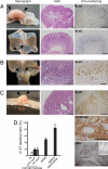
Hematological abnormalities in cagAHs mice. (A) Myeloid leukemia developed in a 72-week-old CAG-cagAHs heterozygous male mouse (B-10). (Upper) Macroscopic and histological views of the spleen and bone marrow (BM) from the mouse with leukemia. Age-matched control B6 spleen is also shown. (Scale bars, 100 μm.) (Lower) FACS analyses of spleen and BM cells from the mice with leukemias. Cells were double-stained with anti-Gr-1 and anti-Mac-1 antibodies, showing increased numbers of Mac-1/Gr-1-double-positive myeloid cells. The percentage of each cell population is indicated. BM cells from B6 mice were used as a control. (B) B cell lymphoma of mesenteric lymph-node origin developed in a 72-week-old CAG-cagAHs (B-10) heterozygous male mice. Macroscopic view, H&E staining, and anti-B220 immunostaining showing diffuse infiltration of B cells into the spleen and liver, resembling diffuse large B cell lymphoma. (Scale bars, 100 μm.) (C) T cell lymphoma developed in a 72-week-old CAG-cagAHs (B-10) homozygous male mice. Macroscopic view, H&E staining and anti-CD3 immunostaining showing infiltration of T cells into the spleen, liver, and lung. (Scale bars, 300 μm.) (D) Blood smears from 72-week-old B6, CAG-cagAHs (B-10) and HK-cagAHs (F-11) heterozygous male mice, showing increased granulocytes in cagAHs mice. (Scale bars, 30 μm.) (E) Immunoblot analysis of Erk phosphorylation in spleen cells from B6 or CAG-cagAHs (B-10) mice. (F) Myeloid colonies derived from bone marrow cells of 72-week-old B6 or CAG-cagAHs (B-10) heterozygous male mice with no evidence of hematopoietic malignancies. Error bars indicate mean ± SD of triplicates. *, P < 0.05, Student's t test.
Fig. 4.
Fig. 4.
Analysis of transgenic mice expressing phospho-resistant (PR) CagA. (A) PR-cagAHs mRNAs in the stomachs of 12-week-old PR-cagAHs heterozygous male mice as determined by RT-PCR analysis. (B) Relative cagAHs mRNA levels in the stomachs of 12-week-old B6, CAG-cagAHs (B-10), HK-cagAHs (A-21), CAG-PR-cagAHs (A-20), and HK-PR-cagAHs (D-01) heterozygous male mice. Error bars indicate mean ± SD of triplicates. (C) Immunoblot analysis of CagA expression in the stomachs of 12-week-old B6, CAG-cagAHs (B-10), HK-cagAHs (A-21), CAG-PR-cagAHs (A-20), and HK-PR-cagAHs (D-01) heterozygous male mice. (D) H&E staining and anti-PCNA immunostaining of the glandular stomachs from 12-week-old B6, CAG-PR-cagAHs (A-20), HK-PR-cagAHs (D-01), and CAG-cagAHs (B-10) heterozygous male mice. *, PCNA-labeled cells. (Scale bars, 300 μm.) Gastric mucosal thicknesses of these mice are shown at Right. **, P > 0.05, Student's t test. (E) Blood smears from 72-week-old B6, CAG-PR-cagAHs (A-20), and HK-PR-cagAHs (B-20) heterozygous male mice. (F) Myeloid colonies derived from bone marrow cells of 72-week-old B6 or CAG-PR-cagAHs (A-20) heterozygous male mice with no evidence of hematopoietic malignancies. Error bars indicate mean ± SD of triplicates. *, P > 0.05, Student's t test.

References

    1. Parkin DM. Global cancer statistics in the year 2000. Lancet Oncol. 2001;2:533–543. - PubMed
    1. Peek RM, Jr, Blaser MJ. Helicobacter pylori and gastrointestinal tract adenocarcinomas. Nat Rev Cancer. 2002;2:28–37. - PubMed
    1. Parsonnet J, et al. Helicobacter pylori infection and gastric lymphoma. N Engl J Med. 1994;330:1267–1271. - PubMed
    1. Kuipers EJ, Perez-Perez GI, Meuwissen SG, Blaser MJ. Helicobacter pylori and atrophic gastritis: importance of the cagA status. J Natl Cancer Inst. 1995;87:1777–1780. - PubMed
    1. Sozzi M, et al. Atrophic gastritis and intestinal metaplasia in Helicobacter pylori infection: the role of CagA status. Am J Gastroenterol. 1998;93:375–379. - PubMed

Publication types

MeSH terms