. 2008 Mar;294(3):L582-91.
doi: 10.1152/ajplung.00353.2007. Epub 2008 Jan 11.

Protein kinase Cdelta regulates endothelial nitric oxide synthase expression via Akt activation and nitric oxide generation

Affiliations

Protein kinase Cdelta regulates endothelial nitric oxide synthase expression via Akt activation and nitric oxide generation

Neetu Sud et al. Am J Physiol Lung Cell Mol Physiol. 2008 Mar.

Retraction in

Abstract

In this study, we explore the roles of the delta isoform of PKC (PKCdelta) in the regulation of endothelial nitric oxide synthase (eNOS) activity in pulmonary arterial endothelial cells isolated from fetal lambs (FPAECs). Pharmacological inhibition of PKCdelta with either rottlerin or with the peptide, deltaV1-1, acutely attenuated NO production, and this was associated with a decrease in phosphorylation of eNOS at Ser1177 (S1177). The chronic effects of PKCdelta inhibition using either rottlerin or the overexpression of a dominant negative PKCdelta mutant included the downregulation of eNOS gene expression that was manifested by a decrease in both eNOS promoter activity and protein expression after 24 h of treatment. We also found that PKCdelta inhibition blunted Akt activation as observed by a reduction in phosphorylated Akt at position Ser473. Thus, we conclude that PKCdelta is actively involved in the activation of Akt. To determine the effect of Akt on eNOS signaling, we overexpressed a dominant negative mutant of Akt and determined its effect of NO generation, eNOS expression, and phosphorylation of eNOS at S1177. Our results demonstrated that Akt inhibition was associated with decreased NO production that correlated with reduced phosphorylation of eNOS at S1177, and decreased eNOS promoter activity. We next evaluated the effect of endogenously produced NO on eNOS expression by incubating FPAECs with the eNOS inhibitor 2-ethyl-2-thiopseudourea (ETU). ETU significantly inhibited NO production, eNOS promoter activity, and eNOS protein levels. Together, our data indicate involvement of PKCdelta-mediated Akt activation and NO generation in maintaining eNOS expression.


Figures

Fig. 1.
Fig. 1.
Acute effects of PKCδ inhibition on Akt activation in pulmonary arterial endothelial cells isolated from fetal lambs (FPAECs). FPAECs were acutely exposed to the PKCδ inhibitor, rottlerin (10 μM, 30 min), the PKCδ-derived inhibitory peptide δV1-1 (1 μM, 2 h), or control peptide (1 μM, 2 h), and then whole cell lysates were subjected to Western blot analysis for total and phopsho-Tyr311 PKCδ (AD) and Akt phospho-Ser473 Akt (EH). Protein loading was also normalized for loading using β-actin. Representative images are shown for each. Although total PKCδ (A and C) and Akt (E and G) were unchanged by PKCδ inhibition, both phopsho-Tyr311 PKCδ (B and D) and phospho-Ser473 Akt (F and H) were significantly decreased. Data are presented as means ± SE, n = 3. *P < 0.05 vs. control cells.
Fig. 2.
Fig. 2.
Acute effects of PKCδ inhibition on NO signaling in pulmonary arterial endothelial cells isolated from fetal lambs. FPAECs were acutely exposed to the PKCδ inhibitor, rottlerin (10 μM, 30 min), the PKCδ-derived inhibitory peptide δV1-1 (1 μM, 2 h), or control peptide (1 μM, 2 h), and then whole cell lysates were subjected to Western blot analysis for eNOS as well as phospho-Ser1177 and phospho-Thr495 eNOS (AD). Protein loading was also normalized for loading using β-actin. Representative images are shown for each. Although total eNOS levels were unchanged by PKCδ inhibition, phospho- phospho-Ser1177 eNOS (D) was significantly decreased. However, phospho-Thr495 eNOS was unchanged (D). The decrease in Ser1177 phosphorylation of eNOS was associated with a significant decrease in NO generation (E and F). Data are presented as means ± SE, n = 3. *P < 0.05 vs. control cells.
Fig. 3.
Fig. 3.
Generation of a PKCδ dominant negative mutant expression plasmid. FPAECs were transfected or not with the dominant negative PKCδ mutant plasmid, pIRES-DNPKCδ. After 24 h, whole cell lysates were subjected to Western blot analysis for the FLAG epitope (A) as well as total and phospho-Tyr311 PKCδ (B and C). Representative images are shown for each. Although total PKCδ (B and C) is unchanged by pIRES-DNPKCδ transfection phospho-Tyr311 PKCδ (C) levels were significantly decreased. Data are presented as means ± SE, n = 3. *P < 0.05 vs. control cells.
Fig. 4.
Fig. 4.
Prolonged PKCδ inhibition decreases eNOS expression in pulmonary arterial endothelial cells isolated from fetal lambs. FPAECs were exposed to the PKCδ inhibitor, rottlerin (10 μM), or transfected with the dominant negative PKCδ mutant, pIRES-DNPKCδ. After 24 h, whole cell lysates were subjected to Western blot analysis to determine the effect on eNOS protein levels. eNOS expression was also normalized for loading using β-actin. Representative images are shown for rottlerin (A) and pIRES-DNPKCδ (C). There is a significant decrease in eNOS expression after 24 h of PKCδ inhibition by both rottlerin (B) and pIRES-DNPKCδ (D). The data are expressed as means ± SE, n = 3. *P < 0.05 vs. control cells.
Fig. 5.
Fig. 5.
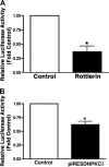
Transcriptional activity of the human eNOS promoter in response to PKCδ inhibition in pulmonary arterial endothelial cells isolated from fetal lambs. FPAECs were transfected with 1.6 kb of upstream sequence of the eNOS promoter fused to a luciferase reporter gene. Cells were also cotransfected with a construct expressing a β-galactosidase reporter gene as a transfection efficiency control. Cells were then either treated with rottlerin (10 μM) or transfected with the dominant negative PKCδ mutant, pIRES-DNPKCδ. After 24 h, luciferase activity was determined. PKCδ inhibition with either rottlerin (A) or pIRES-DNPKCδ (B) significantly decreases the activity of the 1.6-kb human eNOS promoter fragment. Values expressed are means ± SE, n = 6. *P < 0.05 vs. control cells.
Fig. 6.
Fig. 6.
Effect of Akt inhibition on eNOS expression and activity in pulmonary arterial endothelial cells isolated from fetal lambs. FPAECs were transduced with adenoviruses expressing either a dominant negative mutant of Akt or green fluorescent protein (GFP) (as a transduction control). Western blot analysis in whole cell lysates was used to confirm Akt overexpression (A). There is a significant increase in Akt expression with the transduction of the dominant Akt adenovirus (B). The effect of Akt inhibition on phospho-Ser1177 eNOS was then determined by Western blot analysis (C). There is a significant decrease in phospho-Ser1177 with the transduction of the dominant Akt adenovirus (D). Representative images are shown, and in all cases, protein loading was normalized for loading using β-actin. The decrease in Ser1177 phosphorylation of eNOS was also associated with a significant decrease in NO generation (E). Data are presented as means ± SE, n = 3. *P < 0.05 vs. control cells.
Fig. 7.
Fig. 7.
Transcriptional activity of the human eNOS promoter in response to Akt inhibition in pulmonary arterial endothelial cells isolated from fetal lambs. FPAECs were transduced with adenoviruses expressing either a dominant negative mutant of Akt or GFP (as a transduction control). Then, after 24 h, there was further transfection with 1.6-kb of upstream sequence of the eNOS promoter fused to a luciferase reporter gene along with a construct expressing a β-galactosidase reporter gene (as a transfection efficiency control). Cells were harvested after a further 24 h, and the luciferase activity was determined. Akt inhibition significantly decreases the activity of the 1.6-kb human eNOS promoter fragment. Values expressed are means ± SE, n = 6. *P < 0.05 vs. control cells.
Fig. 8.
Fig. 8.
Effect of endogenous NO generation on eNOS protein levels in pulmonary arterial endothelial cells isolated from fetal lambs. FPAECs were exposed to the NOS inhibitor 2-ethyl-2-thiopseudourea (ETU) (100 μM, 24 h). NOS inhibition was confirmed by a significant decrease in NO generation (A). FPAECs were then transfected with 1.6 kb of upstream sequence of the eNOS promoter fused to a luciferase reporter gene. Cells were also cotransfected with a construct expressing a β-galactosidase reporter gene as a transfection efficiency control. Cells were then treated with ETU (100 μM, 24 h), and the luciferase activity was determined. NOS inhibition significantly decreases the activity of the 1.6-kb human eNOS promoter fragment (B). Values expressed are means ± SE, n = 6. *P < 0.05 vs. control cells. Furthermore, to examine effects of NOS inhibition on eNOS protein levels, whole cell lysates were subjected to Western blot analysis. eNOS expression was also normalized for loading using β-actin. A representative image is shown (C). There is a significant decrease in eNOS expression after 24 h of NOS inhibition (D). The data are expressed as means ± SE, n = 3. *P < 0.05 vs. control cells.
Fig. 9.
Fig. 9.
Sodium nitroprusside (SNP) prevents the decrease in eNOS expression in response to NOS inhibition in pulmonary arterial endothelial cells isolated from fetal lambs. FPAECs were transfected with 1.6 kb of upstream sequence of the eNOS promoter fused to a luciferase reporter gene. Cells were also cotransfected with a construct expressing a β-galactosidase reporter gene as a transfection efficiency control. Cells were then treated with ETU (100 μM) in the presence or absence of the NO donor SNP (1 mM). After 24 h, luciferase activity was determined. The significant decrease in eNOS expression induced by NOS inhibition is attenuated by SNP (A). Values expressed are means ± SE, n = 6. *P < 0.05 vs. control cells. Whole cell lysates were also subjected to Western blot analysis to examine eNOS protein levels. Expression was also normalized for loading using β-actin. A representative image is shown (B). The significant decrease in eNOS expression associated with NOS inhibition is attenuated by SNP (C). The data are expressed as means ± SE, n = 3. *P < 0.05 vs. control cells; †P < 0.05 vs. ETU alone.

Similar articles

Cited by

References

    1. Ahn BK, Jeong SK, Kim HS, Choi KJ, Seo JT, Choi EH, Ahn SK, Lee SH. Rottlerin, a specific inhibitor of protein kinase C-delta, impedes barrier repair response by increasing intracellular free calcium. J Invest Dermatol 126: 1348–1355, 2006 - PubMed
    1. Boo YC, Jo H. Flow-dependent regulation of endothelial nitric oxide synthase: role of protein kinases. Am J Physiol Cell Physiol 285: C499–C508, 2003 - PubMed
    1. Boo YC, Sorescu G, Boyd N, Shiojima I, Walsh K, Du J, Jo H. Shear stress stimulates phosphorylation of endothelial nitric-oxide synthase at Ser1179 by Akt-independent mechanisms: role of protein kinase A. J Biol Chem 277: 3388–3396, 2002 - PubMed
    1. Bright R, Raval AP, Dembner JM, Perez-Pinzon MA, Steinberg GK, Yenari MA, Mochly-Rosen D. Protein kinase C delta mediates cerebral reperfusion injury in vivo. J Neurosci 24: 6880–6888, 2004 - PMC - PubMed
    1. Bright R, Steinberg GK, Mochly-Rosen D. DeltaPKC mediates microcerebrovascular dysfunction in acute ischemia and in chronic hypertensive stress in vivo. Brain Res 1144: 146–155, 2007 - PMC - PubMed

Publication types

MeSH terms

LinkOut - more resources