. 2008 Feb;10(2):149-59.
doi: 10.1038/ncb1677. Epub 2008 Jan 13.

Intra-axonal translation and retrograde trafficking of CREB promotes neuronal survival

Affiliations

Intra-axonal translation and retrograde trafficking of CREB promotes neuronal survival

Llewellyn J Cox et al. Nat Cell Biol. 2008 Feb.

Erratum in

  • Nat Cell Biol. 2008 Mar;10(3):370

Abstract

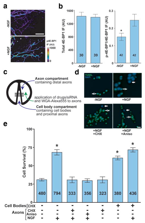
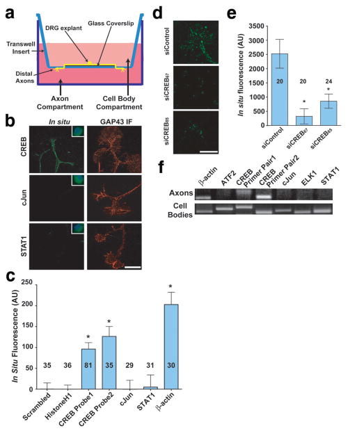
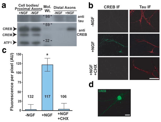
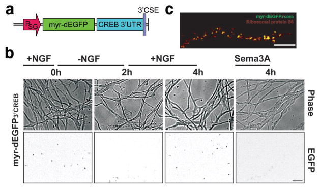
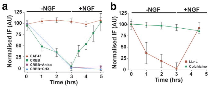
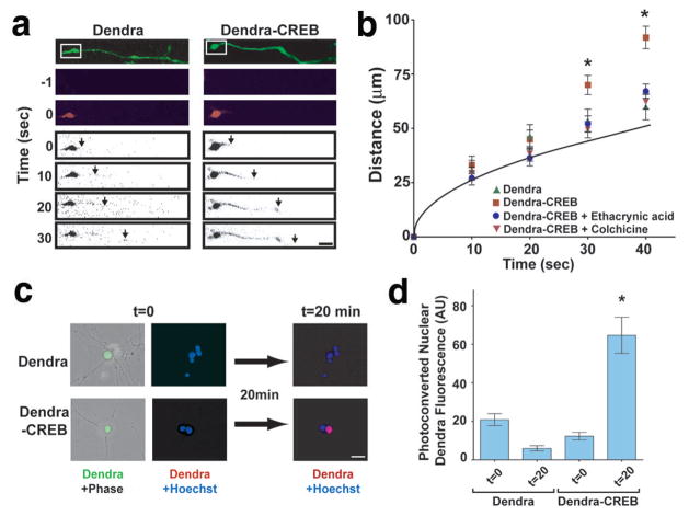
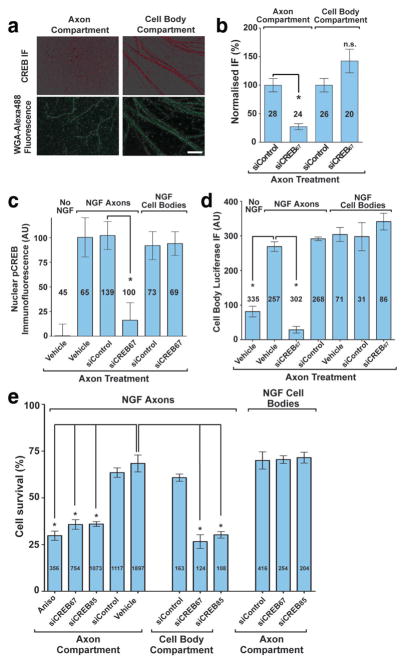
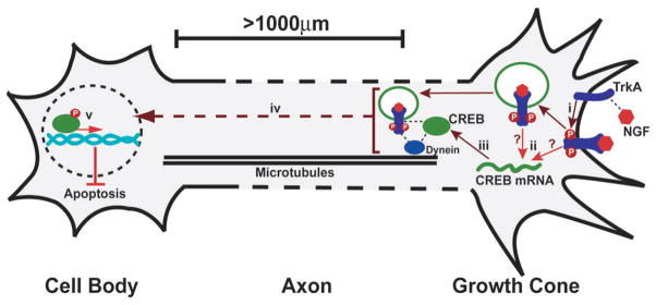
During development of the nervous system, axons and growth cones contain mRNAs such as beta-actin, cofilin and RhoA, which are locally translated in response to guidance cues. Intra-axonal translation of these mRNAs results in local morphological responses; however, other functions of intra-axonal mRNA translation remain unknown. Here, we show that axons of developing mammalian neurons contain mRNA encoding the cAMP-responsive element (CRE)-binding protein (CREB). CREB is translated within axons in response to nerve growth factor (NGF) and is retrogradely trafficked to the cell body. In neurons that are selectively deficient in axonal CREB transcripts, increases in nuclear pCREB, CRE-mediated transcription and neuronal survival elicited by axonal application of NGF are abolished, indicating a signalling function for axonally synthesized CREB. These studies identify a signalling role for axonally derived CREB, and indicate that signal-dependent synthesis and retrograde trafficking of transcription factors enables specific transcriptional responses to signalling events at distal axons.


Figures

Figure 1
Figure 1. Local protein synthesis in axons is required for NGF-dependent survival
(a) Phospho-4E-BP1 levels increase in growth cones in response to NGF treatment. DRG neurons were incubated with NGF-replete or NGF-free media; phospho-4E-BP1 levels in axons were measured by immunofluorescence. Scale bar, 50 μm. (b) Quantification of total and phospho-4EBP1 in (a) p=0.012. Numbers on bars represent n axons per condition. (c) Schematic diagram of compartmented (Campenot) chambers. E15 dissociated DRG neurons are cultured in the cell body compartment and axons grow under a thinly applied silicone grease layer that seals the chamber with the Permanox® plastic culture slide. (d) Application of protein synthesis inhibitors to axons blocks NGF-mediated retrograde survival. Dissociated DRG neurons were grown in compartmentalised chambers, and vehicle or NGF was added to the axon compartment. 1 μM cycloheximide (CHX) or 40 μM anisomycin (Aniso) were added to the axon compartment concurrently with NGF media. Cell body compartments were kept NGF-free during the course of the experiment. Cells crossing the divider were retrogradely labelled with WGA-Alexa555 and only WGA-positive cell bodies were counted in the data set. Survival was assessed by TUNEL assay. Examples of non-apoptotic and apoptotic are indicated with closed and open arrows, respectively (Blue=DAPI, Green=TUNEL). Scale bar, 20 μm. (e) Quantification of results from (d). *p<0.001. Numbers on bars represent n cells per condition.
Figure 2
Figure 2. CREB mRNA and protein are localised to developing axons of DRG neurons
(a) Schematic diagram of Boyden chamber. DRGs were cultured in the centre of a glass coverslip placed on top of the microporous membrane. Axons grow across the coverslip and cross through the membrane by DIV4, when they are subjected to experimental conditions and mechanically harvested for analysis. (b) Fluorescent in situ hybridisation (FISH) using riboprobes demonstrated the presence of CREB, but not cJun or STAT1 mRNA transcripts in axons. Insets show labelling in cell bodies of dissociated DRG neuron cultures at 10X magnification to demonstrate efficacy of riboprobes. Counter-stained images show immunofluorescence using anti-GAP-43 antibody (Red). Scale bar, 10 μm. (c) Quantification of FISH data in (b). CREB levels were monitored with two separate probes (Table S1), and CREB levels were comparable to those of β-actin. Background FISH levels were defined as the average signal obtained using a scrambled β-actin riboprobe and subtracted from all other data. *p<0.001. Numbers on bars represent n axons per condition. (d) DRG neurons transfected with non-targeting siRNA demonstrated CREB mRNA FISH signals in axons, while neurons transfected with CREB-specific siRNA demonstrated a near-complete abolishment of CREB mRNA FISH signal. Scale bar, 10 μm. (e) Quantification of data in (d). *p<0.001. Numbers on bars represent n axons per condition. (f) CREB was detected in axonal lysates by RT-PCR using two separate primer pairs (Table S2). RT-PCR fidelity was assayed by concurrent RT-PCR from DRG cell body lysates.
Figure 3
Figure 3. CREB is specifically translated in axons
(a) Selective localisation and induction of CREB in distal axons. DRG explants were cultured in Boyden chambers and the upper compartment was incubated in 0ng/ml NGF. The lower compartment was incubated in either 0 ng/ml NGF or 100 ng/ml NGF for 3 h. Lysates (25 μg protein) were prepared from the coverslip (cell body/proximal axon) or the underside of the membrane (distal axons) and analysed by Western blot using an antibody that also recognises CREB family members CREM and ATF-1. (b) NGF and protein synthesis are required for CREB localisation in axon terminals. Axons were severed from DIV3 DRG explant cultures, and incubated with 0 or 100 ng/ml NGF, or 100 ng/ml NGF + 1 μM cycloheximide (CHX) for 3 h. CREB was detected by immunofluorescence (IF) using a CREB-specific antibody. Counter-staining shows immunofluorescence using anti-tau antibody (right). Scale bar, 50 μm. (c) Quantification of results in (b). *p<0.0001. Numbers on bars represent n axons per condition. (d) Low-power (20X) image of CREB immunofluorescence (IF) in DIV3 E15 DRGs. The majority of cellular CREB protein is associated with the nucleus, although signals are seen in the cytosol and axon. Scale bar 20 μm.
Figure 4
Figure 4. NGF dependent translation of a CREB reporter mRNA in axons
(a) Schematic diagram of the Sindbis viral reporter construct used to monitor CREB translation. The reporter contains a myristoylated, destabilised EGFP (myr-dEGFP) with the 3′UTR of CREB, expressed under control of the Sindbis subgenomic promoter (PSG). (b) E15 DRG explant cultures were infected with Sindbis constructs expressing myr-dEGFP3′CREB on DIV3 and fluorescence (bottom panel) and phase (top panel) images, approximately 1000 μm from the cell body were collected after 24 h. Fluorescence images are shown in inverted contrast in order to more readily visualise puncta. Scale bar, 25 μm. (c) myr-dEGFP3′ CREB puncta colocalise with ribosomal markers. myr-dEGFP3′ CREB-expressing axons were counter-stained by immunofluorescence using a ribosomal protein S6-specific antibody. EGFP fluorescence colocalises with a population of S6-labeled ribosomal clusters. Scale bar, 10μm.
Figure 5
Figure 5. Axonal translation and retrograde transport of endogenous CREB
(a) CREB levels in severed axons of E15 DRGs were assayed by immunofluorescence with a CREB-specific antibody. CREB was depleted upon removal of NGF, with near complete loss of fluorescence signals by 3 h. Restoration of NGF resulted in a return of CREB to starting levels within 2 h. Levels of GAP-43 were not significantly affected by NGF removal or by restoration of NGF, indicating that changes in fluorescence intensity were not due to significant changes in axonal volume. Application of 1 μM cycloheximide or 40 μM anisomycin, concurrent with NGF replacement, prevented the restoration of CREB immunofluorescence, indicating that the NGF-dependent increase in CREB levels requires protein synthesis. n≥40 axons per data point. (b) CREB is depleted from axon terminals in a microtubule-dependent manner. CREB levels in severed axons were monitored as in (a) following removal and restoration of NGF, in the presence of LLnL or colchicine. Colchicine, but not LLnL, blocked the reduction in CREB levels following removal of NGF. n≥40 axons per data point.
Figure 6
Figure 6. Retrograde transport of a photoactivatable fluorescent CREB reporter protein
(a) E15 DRG explant cultures were infected with Sindbis constructs expressing Dendra or Dendra-CREB transcripts. Dendra and Dendra-CREB were visualised as green fluorescence (top panel), and growth cone-localised Dendra[-CREB] was photoactivated to its red form by 50 ms illumination of the boxed regions with a 408 nm laser. Movement of photoactivated Dendra signals was analysed within the axon determined by green Dendra fluorescence mask. The leading edge (arrows) of red fluorescence for photoactivated Dendra-CREB was observed to move along the axon at a significantly faster rate than photoactivated Dendra. Photoactivated Dendra[-CREB] images are shown inverted in order to more readily visualise signals. Scale bar, 10 μm. (b) Quantification of data in (a). Grey line indicates the predicted diffusion rate of photoconverted Dendra-CREB, based on neuronal viscosity measurements. The expected diffusion rate of Dendra-CREB was calculated at various elapsed time points, using a previously measured diffusion coefficient (D) in neurons, in the formula x2 = (2Dt), where x is the average displacement. No significant differences in axon diameter or morphology were observed between the neurons assayed. n≥20 axons per data point. *p<0.0001. (c) Dendra or Dendra-CREB was photoactivated by 1 s illumination of a 40 μm axon segment approximately 1 mm from its respective cell body and levels of photoactivated Dendra fluorescence were analysed in the respective cell nucleus. Scale bar, 20 μm. (d) Quantification of data in (c). n=10 cells per data point. *p=0.0012.
Figure 7
Figure 7. Axonal CREB is required for CRE-dependent transcription and NGF-mediated DRG survival
(a) DRG axons in compartmented cultures were transfected with CREB siRNA in the axon compartment only, and CREB levels were detected using a CREB antibody. Axons crossing the compartment divider were retrogradely labelled with WGA-Alexa488. All compartments were maintained in NGF-containing media throughout the experiment. Scale bar, 50 μm. (b) Quantification of data in (a). Immunofluorescence levels in each compartment were normalised to fluorescence signals from cultures treated with non-targeting siRNA. *p<0.0001. Numbers on bars represent n axons per condition. (c) DRGs in compartmented cultures were incubated in NGF-free media, supplemented with BAF to suppress apoptosis. Axon compartments were treated with CREB-specific or non-targeting siRNA. After 48 h, 30 ng/ml NGF was added to the axon compartment for 20 min, after which pCREB levels in nuclei were quantified by immunofluorescence. *p=0.0004. Numbers on bars represent n cells per condition. (d) DRGs in compartmented chambers were infected with adenovirus encoding luciferase under the control of a CRE transcriptional element and treated with NGF as in (c). *p<0.001. Numbers on bars represent n cells per condition. (e) E15 dissociated DRG were cultured in compartmented chambers as in Fig. 1c. NGF-induced neuronal survival at DIV7 was assayed following transfection of control or CREB-specific siRNA into the axon compartment at DIV5. *p<0.001. Numbers on bars represent n cells per condition.
Figure 8
Figure 8. Local translation and retrograde transport of CREB mediates neuronal survival
(i) NGF binds to TrkA receptors causing dimerisation and autophosphorylation. (ii) TrkA activation leads to translation of axonal CREB mRNA and (iii) the production of CREB protein. NGF-bound, activated TrkA receptors are internalised into endosomes and initiate formation of a signalling complex containing downstream effectors and the motor protein dynein. Axonally translated CREB protein associates with this NGF-pTrkA signalling endosome, which is required for downstream activation of CREB signalling in the cell body. (iv) CREB is retrogradely transported to the nucleus via microtubules and is phosphorylated at S133 downstream of internalised TrkA signalling endosomes, via a kinase cascade including Mek5/Erk5. (v) Axonally-derived pCREB initiates the transcription of anti-apoptotic genes in the nucleus, leading to neuronal cell survival.

Comment in

References

    1. Czaplinski K, Singer RH. Pathways for mRNA localization in the cytoplasm. Trends Biochem Sci. 2006;31:687–693. - PubMed
    1. Piper M, Holt C. RNA translation in axons. Annual Review of Cell and Developmental Biology. 2004;20:505–523. - PMC - PubMed
    1. Bassell GJ, Zhang H, Byrd AL, Femino AM, Singer RH, Taneja KL, Lifshitz LM, Herman IM, Kosik KS. Sorting of β-actin mRNA and protein to neurites and growth cones in culture. Journal of Neuroscience. 1998;18:251–265. - PMC - PubMed
    1. Wu K, Hengst U, Cox LJ, Macosko EZ, Jeromin A, Urquhart ER, Jaffrey SR. Local translation of RhoA regulates growth cone collapse. Nature. 2005;436:1020–1024. - PMC - PubMed
    1. Piper M, Anderson R, Dwivedy A, Weinl C, van Horck F, Leung KM, Cogill E, Holt C. Signaling mechanisms underlying Slit2-induced collapse of Xenopus retinal growth cones. Neuron. 2006;49:215–228. - PMC - PubMed

Publication types

Substances