

Kinetics of G-protein-coupled receptor signals in intact cells
- PMID: 18193071
- PMCID: PMC2268076
- DOI: 10.1038/sj.bjp.0707656
Kinetics of G-protein-coupled receptor signals in intact cells
Abstract
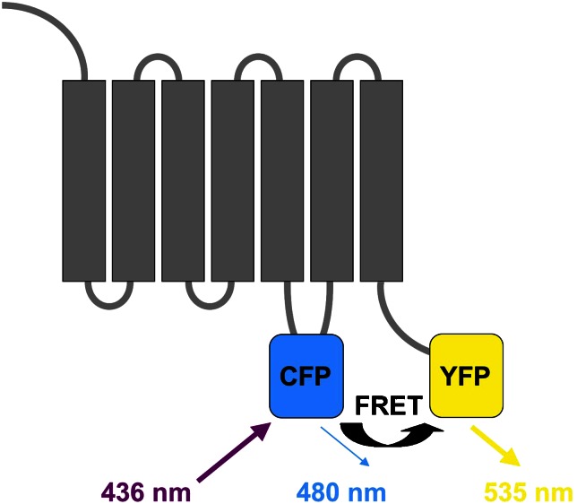
G-protein-coupled receptors (GPCRs) are the largest group of cell surface receptors. They are stimulated by a variety of stimuli and signal to different classes of effectors, including several types of ion channels and second messenger-generating enzymes. Recent technical advances, most importantly in the optical recording with energy transfer techniques--fluorescence and bioluminescence resonance energy transfer, FRET and BRET--, have permitted a detailed kinetic analysis of the individual steps of the signalling chain, ranging from ligand binding to the production of second messengers in intact cells. The transfer of information, which is initiated by ligand binding, triggers a signalling cascade that displays various rate-controlling steps at different levels. This review summarizes recent findings illustrating the speed and the complexity of this signalling system.
Figures
References
-
- Azpiazu I, Gautam N. A fluorescence resonance energy transfer-based sensor indicates that receptor access to a G protein is unrestricted in a living mammalian cell. J Biol Chem. 2004;279:27709–27718. - PubMed
-
- Bourne HR, Sanders DA, McCormick F. The GTPase superfamily: conserved structure and molecular mechanism. Nature. 1991;349:117–127. - PubMed
-
- Bünemann M, Bücheler MM, Philipp M, Lohse MJ, Hein L. Activation and deactivation kinetics of α2A- and α2C-adrenergic receptor-activated G protein-activated inwardly rectifying K+-channel currents. J Biol Chem. 2001;276:47512–47517. - PubMed
Publication types
MeSH terms
Substances
LinkOut - more resources
Full Text Sources
