Detrimental role of endogenous nitric oxide in host defence against Sporothrix schenckii
- PMID: 18194265
- PMCID: PMC2433314
- DOI: 10.1111/j.1365-2567.2007.02712.x
Detrimental role of endogenous nitric oxide in host defence against Sporothrix schenckii
Abstract
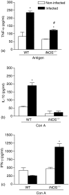
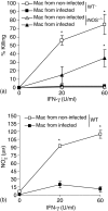
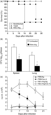
We earlier demonstrated that nitric oxide (NO) is a fungicidal molecule against Sporothrix schenckii in vitro. In the present study we used mice deficient in inducible nitric oxide synthase (iNOS-/-) and C57BL/6 wild-type (WT) mice treated with Nomega-nitro-arginine (Nitro-Arg-treated mice), an NOS inhibitor, both defective in the production of reactive nitrogen intermediates, to investigate the role of endogenous NO during systemic sporotrichosis. When inoculated with yeast cells of S. schenckii, WT mice presented T-cell suppression and high tissue fungal dissemination, succumbing to infection. Furthermore, susceptibility of mice seems to be related to apoptosis and high interleukin-10 and tumour necrosis factor-alpha production by spleen cells. In addition, fungicidal activity and NO production by interferon-gamma (IFN-gamma) and lipopolysaccharide-activated macrophages from WT mice were abolished after fungal infection. Strikingly, iNOS-/- and Nitro-Arg-treated mice presented fungal resistance, controlling fungal load in tissues and restoring T-cell activity, as well as producing high amounts of IFN-gamma Interestingly, macrophages from these groups of mice presented fungicidal activity after in vitro stimulation with higher doses of IFN-gamma. Herein, these results suggest that although NO was an essential mediator to the in vitro killing of S. schenckii by macrophages, the activation of NO system in vivo contributes to the immunosuppression and cytokine balance during early phases of infection with S. schenckii.
Figures


Similar articles
-
Response of macrophage Toll-like receptor 4 to a Sporothrix schenckii lipid extract during experimental sporotrichosis.Immunology. 2009 Oct;128(2):301-9. doi: 10.1111/j.1365-2567.2009.03118.x. Immunology. 2009. PMID: 19740386 Free PMC article.
-
Dectin-1 expression by macrophages and related antifungal mechanisms in a murine model of Sporothrix schenckii sensu stricto systemic infection.Microb Pathog. 2017 Sep;110:78-84. doi: 10.1016/j.micpath.2017.06.025. Epub 2017 Jun 20. Microb Pathog. 2017. PMID: 28645771
-
Influence of TLR-2 in the immune response in the infection induced by fungus Sporothrix schenckii.Immunol Invest. 2014;43(4):370-90. doi: 10.3109/08820139.2013.879174. Epub 2014 Jan 31. Immunol Invest. 2014. PMID: 24484374
-
Cell wall proteins of Sporothrix schenckii as immunoprotective agents.Rev Iberoam Micol. 2014 Jan-Mar;31(1):86-9. doi: 10.1016/j.riam.2013.09.017. Epub 2013 Nov 17. Rev Iberoam Micol. 2014. PMID: 24257472 Review.
-
The immune response against Candida spp. and Sporothrix schenckii.Rev Iberoam Micol. 2014 Jan-Mar;31(1):62-6. doi: 10.1016/j.riam.2013.09.015. Epub 2013 Nov 16. Rev Iberoam Micol. 2014. PMID: 24252829 Review.
Cited by
-
Increased leptin response and inhibition of apoptosis in thymocytes of young rats offspring from protein deprived dams during lactation.PLoS One. 2013 May 10;8(5):e64220. doi: 10.1371/journal.pone.0064220. Print 2013. PLoS One. 2013. PMID: 23675529 Free PMC article.
-
Nitric oxide synthase activity has limited influence on the control of Coccidioides infection in mice.Microb Pathog. 2011 Sep;51(3):161-8. doi: 10.1016/j.micpath.2011.03.013. Epub 2011 Apr 14. Microb Pathog. 2011. PMID: 21513788 Free PMC article.
-
Disseminated sporotrichosis.BMJ Case Rep. 2011 Mar 25;2011:bcr1020103404. doi: 10.1136/bcr.10.2010.3404. BMJ Case Rep. 2011. PMID: 22700076 Free PMC article.
-
Response of macrophage Toll-like receptor 4 to a Sporothrix schenckii lipid extract during experimental sporotrichosis.Immunology. 2009 Oct;128(2):301-9. doi: 10.1111/j.1365-2567.2009.03118.x. Immunology. 2009. PMID: 19740386 Free PMC article.
-
Involvement of major components from Sporothrix schenckii cell wall in the caspase-1 activation, nitric oxide and cytokines production during experimental sporotrichosis.Mycopathologia. 2015 Feb;179(1-2):21-30. doi: 10.1007/s11046-014-9810-0. Epub 2014 Sep 10. Mycopathologia. 2015. PMID: 25205196
References
-
- Morris-Jones R. Sporotrichosis. Clin Exp Dermatol. 2002;27:427–31. - PubMed
-
- Lopes-Bezerra LM, Schubach A, Costa RO. Sporothrix schenckii and sporotrichosis. Ann Braz Acad Sci. 2006;78:1–16. - PubMed
-
- Aarestrup FM, Guerra RO, Vieira BJ, Cunha RM. Oral manifestation of sporotrichosis in AIDS patients. Oral Dis. 2001;7:134–6. - PubMed
-
- Carvalho MT, de Castro AP, Baby C, Werner B, Filus Neto J, Queiroz-Telles F. Disseminated cutaneous sporotrichosis in a patient with AIDS. report of a case. Rev Soc Bras Med Trop. 2002;35:655–9. - PubMed
Publication types
MeSH terms
Substances
LinkOut - more resources
Full Text Sources
Molecular Biology Databases

