

Matrix vesicles in the fibrous cap of atherosclerotic plaque: possible contribution to plaque rupture
- PMID: 18194456
- PMCID: PMC4506172
- DOI: 10.1111/j.1582-4934.2008.00230.x
Matrix vesicles in the fibrous cap of atherosclerotic plaque: possible contribution to plaque rupture
Abstract
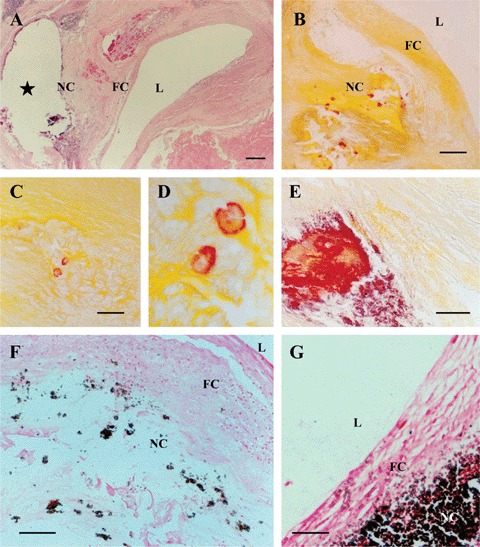
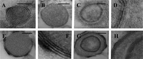
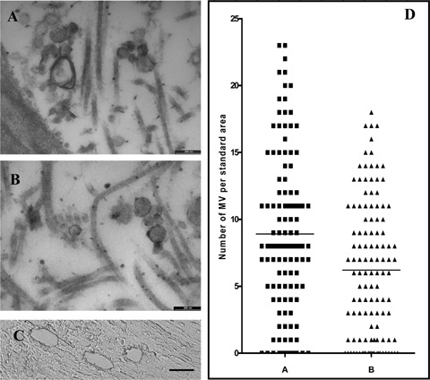
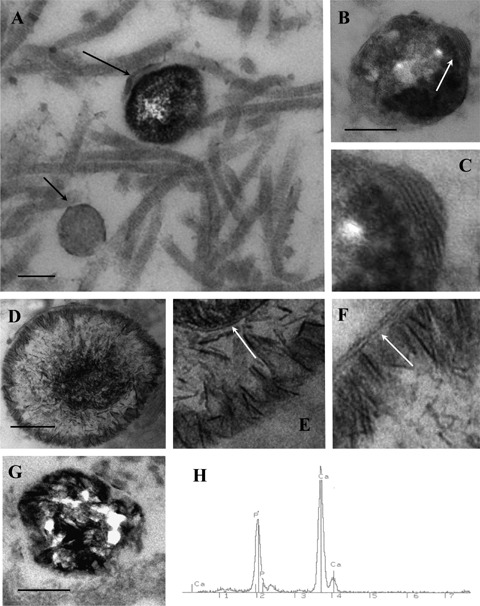
Plaque rupture is the most common type of plaque complication and leads to acute ischaemic events such as myocardial infarction and stroke. Calcification has been suggested as a possible indicator of plaque instability. Although the role of matrix vesicles in the initial stages of arterial calcification has been recognized, no studies have yet been carried out to examine a possible role of matrix vesicles in plaque destabilization. Tissue specimens selected for the present study represented carotid specimens obtained from patients undergoing carotid endarterectomy. Serial frozen cross-sections of the tissue specimens were cut and mounted on glass slides. The thickness of the fibrous cap (FCT) in each advanced atherosclerotic lesion, containing a well developed lipid/necrotic core, was measured at its narrowest sites in sets of serial sections. According to established criteria, atherosclerotic plaque specimens were histologically subdivided into two groups: vulnerable plaques with thin fibrous caps (FCT <100 microm) and presumably stable plaques, in which fibrous caps were thicker than 100 microm. Twenty-four carotid plaques (12 vulnerable and 12 presumably stable plaques) were collected for the present analysis of matrix vesicles in fibrous caps. In order to provide a sufficient number of representative areas from each plaque, laser capture microdissection (LCM) was carried out. The quantification of matrix vesicles in ultrathin sections of vulnerable and stable plaques revealed that the numbers of matrix vesicles were significantly higher in fibrous caps of vulnerable plaques than those in stable plaques (8.908+0.544 versus 6.208+0.467 matrix vesicles per 1.92 microm2 standard area; P= 0.0002). Electron microscopy combined with X-ray elemental microanalysis showed that some matrix vesicles in atherosclerotic plaques were undergoing calcification and were characterized by a high content of calcium and phosphorus. The percentage of calcified matrix vesicles/microcalcifications was significantly higher in fibrous caps in vulnerable plaques compared with that in stable plaques (6.705+/-0.436 versus 5.322+/-0494; P= 0.0474). The findings reinforce a view that the texture of the extracellular matrix in the thinning fibrous cap of atherosclerotic plaque is altered and this might contribute to plaque destabilization.
Figures
References
-
- Burke AP, Farb A, Malcom GT, Liang YH, Smialek J, Virmani R. Coronary risk factors and plaque morphology in men with coronary disease who died suddenly. N EnglJMed. 1997;336:1276–82. - PubMed
-
- Shah PK. Mechanisms of plaque vulnerability and rupture. J Am Coll Cardiol. 2003;41:S15–22. - PubMed
-
- Mitra AK, Dhume AS, Agrawal DK. “Vulnerable plaques’-ticking of the time bomb. Can J Physiol Pharmacol. 2004;82:860–71. - PubMed
-
- Hennerici MG. The unstable plaque. Cerebrovasc Dis. 2004;17(Suppl 3):17–22. - PubMed
-
- Fuster V, Moreno PR, Fayad ZA, Corti R, Badimon JJ. Atherothrombosis and high-risk plaque: part I: evolving concepts. J Am Coll Cardiol. 2005;46:937–54. - PubMed
Publication types
MeSH terms
Substances
LinkOut - more resources
Full Text Sources
Medical
Miscellaneous
