. 2008 Dec;12(6B):2781-9.
doi: 10.1111/j.1582-4934.2008.00215.x. Epub 2008 Jan 11.

The Kazal motifs of RECK protein inhibit MMP-9 secretion and activity and reduce metastasis of lung cancer cells in vitro and in vivo

Affiliations

The Kazal motifs of RECK protein inhibit MMP-9 secretion and activity and reduce metastasis of lung cancer cells in vitro and in vivo

Chong-Keng Chang et al. J Cell Mol Med. 2008 Dec.

Abstract

RECK is a membrane-anchored glycoprotein which may negatively regulate matrix metalloproteinase (MMP) activity to suppress tumor invasion and metastasis. In this study, recombinant proteins corresponding to the residues 285-368 (named as CKM which contained cysteine knot motif), 605-799 (named as K123 which contained three Kazal motifs), 676-799 (named as K23 which contained the last two Kazal motifs) and full-length RECK were produced and their anti-cancer effects were tested. Full-length RECK and K23 but not K123 and CKM inhibited MMP9 secretion and activity. In addition, RECK and K23 inhibited invasion but not migration of metastatic lung cancer cells in vitro. Protein binding and kinetic study indicated that K23 physically interacted with MMP-9 and inhibited its activity by a non-competitive manner. Moreover, K23 reduced metastatic tumor growth in lungs of nude mice. Taken together, our results suggest that the K23 motifs of RECK protein can inhibit MMP-9 secretion and activity and attenuate metastasis of lung cancer cells.


Figures

Fig. 1
Fig. 1
Production of full-length and functional motifs of RECK protein. (A) Full-length RECK expression vector was generated by subcloning human RECK cDNA into the pSecTag2 vector and transfected into 293T cells. Conditioned medium was collected and the secreted recombinant proteins were purified by using a nickel-chelating resin. Western blot analysis was performed to detect Hisx6-tagged RECK protein. (B) K123 expression vector was constructed by amplifying the cDNA by PCR reaction and the amplified DNA fragment was subcloned into pSecTag2 vector for protein production. (C) K23 expression vector was constructed by subcloning the PCR-amplified DNA fragment into pSecTag2 vector for protein production. (D) CKM protein was produced as described above and purified by a nickel-chelating resin. The glycosy-lated forms of CKM protein (Gly-CKM) were also indicated.
Fig. 2
Fig. 2
Inhibition of cell invasion by full-length RECK and K23. (A) A549 cells were incubated with BSA (B) or various recombinant proteins (1 or 3 μg/ml) and placed in the upper part of the transwell unit for migration assay. The number of migrated cells of the group treated with 3 μg/ml of BSA (B) was defined as 100% (n = 3). (B) CL1.5 cells were treated as described above and the effect of various recombinant proteins on cell migration was studied (n = 3). (C) A549 cells in mediums containing different recombinant proteins were placed in the upper part of the transwell unit coated with Matrigel. The number of invaded cells of the control group treated with 3 μg/ml of BSA was defined as 100% (n = 3). *P < 0.05 when the results of recombinant protein-treated groups were compared with the results of control group. (D) CL1.5 cells were subjected to in vitro invasion assay as described above (n = 3). *P < 0.05 when the results of recombinant protein-treated groups were compared with the results of control group.
Fig. 3
Fig. 3
Full-length RECK and K23 inhibited MMP-9 secretion and activity. (A) Conditioned medium of A549 cells was incubated with 3 μg/ml of BSA (B), CKM, K123, K23 or full-length RECK at 37°C for 30 min. The MMP activity was studied as described in Materials and Methods. Results of three independent experiments were expressed as mean ± SE. MMP activity of the control group incubated with 3 μg/ml of BSA was defined as 100% (n = 3). *P < 0.05 when the results of recombinant protein-treated groups were compared with the results of control group. (B) Conditioned medium of CL1.5 cells was also subjected to MMP activity assay as described above (n = 3). *P < 0.05 when the results of recombinant protein-treated groups were compared with the results of control group. (C) BSA and recombinant proteins (3 μg/ml) were added into the culture medium of A549 cells and incubated for 24 hrs. Conditioned medium from an equal number of cells was subjected to gelatin zymography analysis. The positions of pro-MMP-9 and active MMP-9 were indicated. (D) Cells were treated without (−) or with EGF (200 ng/ml) in the absence or presence of K23 (3 μg/ml) for 4 hrs and RT-PCR was performed to investigate MMP-9 mRNA level. (E) Cells were treated as described above and conditioned medium was collected at 24 hrs after treatment. MMP activity of the conditioned medium was studied (n = 3). *P < 0.05 when the results of two experimental groups were compared.
Fig. 4
Fig. 4
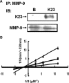
K23 physically interacted with active MMP-9 and functioned as a non-competitive inhibitor. (A) Active recombinant MMP-9 protein (100 ng) was incubated with BSA (B) or K23 (500 ng) at 4°C for 2 hrs. Anti-MMP-9 antibody was added and the immunocomplex was precipitated by protein G-agarose beads. The binding of K23 to MMP-9 was detected by using anti-Hisx6 antibody and the blots were also probed with anti-MMP-9 antibody to confirm that the equal amount of MMP-9 was immunoprecipitated. (B) Active recombinant MMP-9 (5 ng) was mixed with various concentrations of a fluorescein-conjugated gelatin (DQ™ gelatin) and recombinant K23 protein (0, 41.7 or 125 nM). Experiments were performed similar to the procedures of MMP assay as described in Materials and methods.
Fig. 5
Fig. 5
Inhibition of in vivo metastasis of lung cancer cells by K23. (A) A549 stable transfectants expressing pSecTag2 (C) or K23-expressing pSecTag2 (K23) vectors were selected by continuous treatment of hygomycin. Expression of K23 protein was examined by Western blot analysis. (B) The invasive ability of the stable transfectants was investigated as described in Fig. 2C. The invaded cell number of cells transfected with pSecTag2 (C) was defined as 100% (n = 3). *P < 0.05 when the results of K23-expressing group was compared with the results of control group. (C) A total of 1 × 10⁶ stable transfectants were injected into the tail vein of the BALB/c nude mice. Eight weeks after inoculation, animals were sacrificed and the lungs of the animals were inspected for metastatic nodules (indicated by arrow sign). Typical figures of lungs from the two experimental groups were shown. (D) Metastatic nodules were averaged and expressed as Mean ±SE (n = 5). *P < 0.05 when the results of K23-expressing group were compared with the results of control group.

References

    1. Mott JD, Werb Z. Regulation of matrix biology by matrix metalloproteinases. Curr Opin Cell Biol. 2004;16:558–64. - PMC - PubMed
    1. Hojilla CV, Mohammed FF, Khokha R. Matrix metalloproteinases and their tissue inhibitors direct cell fate during cancer development. Br J Cancer. 2003;89:1817–21. - PMC - PubMed
    1. Yan C, Boyd DD. Regulation of matrix metalloproteinase gene expression. J Cell Physiol. 2007;211:19–26. - PubMed
    1. Chakraborti S, Mandal M, Das S, Mandal A, Chakraborti T. Regulation of matrix metalloproteinases: an overview. Mol Cell Biochem. 2003;253:269–85. - PubMed
    1. Visse R, Nagase H. Matrix metallopro-teinases and tissue inhibitors of metallo-proteinases: structure, function, and biochemistry. Circ Res. 2003;92:827–39. - PubMed

Publication types