

Mass spectrometric identification of lysine residues of heme oxygenase-1 that are involved in its interaction with NADPH-cytochrome P450 reductase
- PMID: 18194664
- PMCID: PMC2279806
- DOI: 10.1016/j.bbrc.2008.01.016
Mass spectrometric identification of lysine residues of heme oxygenase-1 that are involved in its interaction with NADPH-cytochrome P450 reductase
Abstract
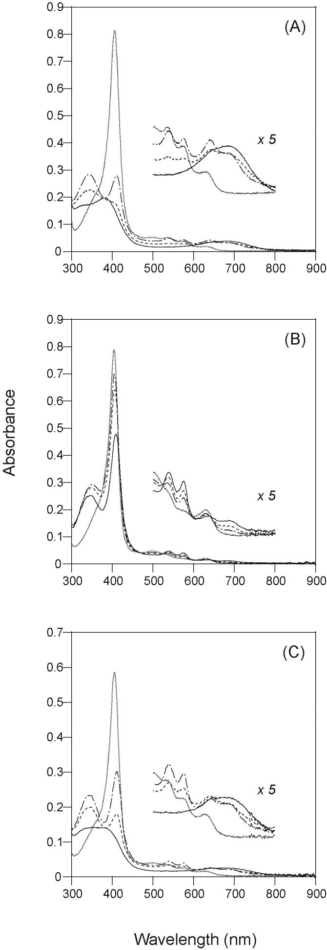
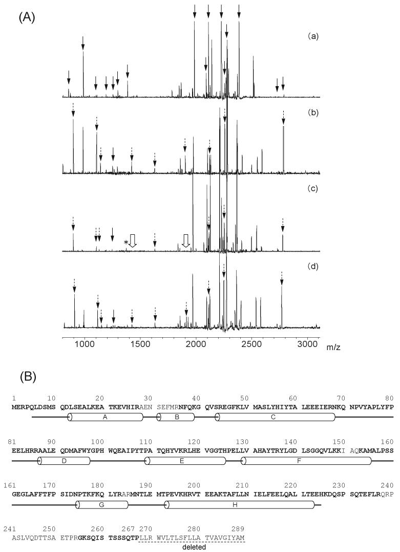
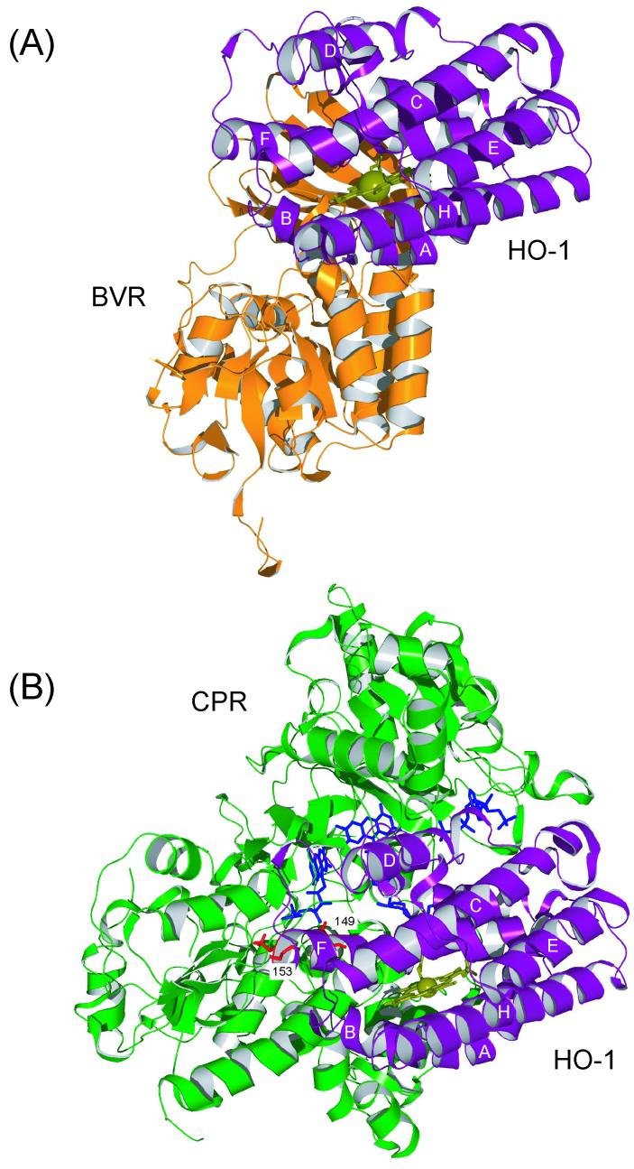
The lysine residues of rat heme oxygenase-1 (HO-1) were acetylated by acetic anhydride in the absence and presence of NADPH-cytochrome P450 reductase (CPR) or biliverdin reductase (BVR). Nine acetylated peptides were identified by MALDI-TOF mass spectrometry in the tryptic fragments obtained from HO-1 acetylated without the reductases (referred to as the fully acetylated HO-1). The presence of CPR prevented HO-1 from acetylation of lysine residues, Lys-149 and Lys-153, located in the F-helix. The heme degradation activity of the fully acetylated HO-1 in the NADPH/CPR-supported system was significantly reduced, whereas almost no inactivation was detected in HO-1 in the presence of CPR, which prevented acetylation of Lys-149 and Lys-153. On the other hand, the presence of BVR showed no protective effect on the acetylation of HO-1. The interaction of HO-1 with CPR or BVR is discussed based on the acetylation pattern and on molecular modeling.
Figures
Similar articles
-
Protein/protein interactions in the mammalian heme degradation pathway: heme oxygenase-2, cytochrome P450 reductase, and biliverdin reductase.J Biol Chem. 2014 Oct 24;289(43):29836-58. doi: 10.1074/jbc.M114.582783. Epub 2014 Sep 7. J Biol Chem. 2014. PMID: 25196843 Free PMC article.
-
The interaction of cytochrome P450 17alpha with NADPH-cytochrome P450 reductase, investigated using chemical modification and MALDI-TOF mass spectrometry.Biochim Biophys Acta. 2006 Jun;1764(6):1126-31. doi: 10.1016/j.bbapap.2006.04.003. Epub 2006 Apr 7. Biochim Biophys Acta. 2006. PMID: 16713412
-
Involvement of NADPH in the interaction between heme oxygenase-1 and cytochrome P450 reductase.J Biol Chem. 2005 Jan 7;280(1):729-37. doi: 10.1074/jbc.M406203200. Epub 2004 Oct 31. J Biol Chem. 2005. PMID: 15516695
-
Recent Advances in the Understanding of the Reaction Chemistries of the Heme Catabolizing Enzymes HO and BVR Based on High Resolution Protein Structures.Curr Med Chem. 2020;27(21):3499-3518. doi: 10.2174/0929867326666181217142715. Curr Med Chem. 2020. PMID: 30556496 Free PMC article. Review.
-
Heme oxygenase and heme degradation.Biochem Biophys Res Commun. 2005 Dec 9;338(1):558-67. doi: 10.1016/j.bbrc.2005.08.020. Epub 2005 Aug 15. Biochem Biophys Res Commun. 2005. PMID: 16115609 Review.
Cited by
-
Oligomerization is crucial for the stability and function of heme oxygenase-1 in the endoplasmic reticulum.J Biol Chem. 2009 Aug 21;284(34):22672-9. doi: 10.1074/jbc.M109.028001. Epub 2009 Jun 25. J Biol Chem. 2009. PMID: 19556236 Free PMC article.
-
Dynamic control of electron transfers in diflavin reductases.Int J Mol Sci. 2012 Nov 15;13(11):15012-41. doi: 10.3390/ijms131115012. Int J Mol Sci. 2012. PMID: 23203109 Free PMC article. Review.
-
Amino Acid Insertion Frequencies Arising from Photoproducts Generated Using Aliphatic Diazirines.J Am Soc Mass Spectrom. 2017 Oct;28(10):2011-2021. doi: 10.1007/s13361-017-1730-z. Epub 2017 Aug 10. J Am Soc Mass Spectrom. 2017. PMID: 28799075
-
Probing protein structure by amino acid-specific covalent labeling and mass spectrometry.Mass Spectrom Rev. 2009 Sep-Oct;28(5):785-815. doi: 10.1002/mas.20203. Mass Spectrom Rev. 2009. PMID: 19016300 Free PMC article. Review.
-
C-Terminal membrane spanning region of human heme oxygenase-1 mediates a time-dependent complex formation with cytochrome P450 reductase.Biochemistry. 2009 Jan 13;48(1):190-7. doi: 10.1021/bi801912z. Biochemistry. 2009. PMID: 19123922 Free PMC article.
References
-
- Tenhunen R, Marver HS, Schmid R. Microsomal heme oxygenase. Characterization of the enzyme. J. Biol. Chem. 1969;244:6388–6394. - PubMed
-
- Yoshida T, Noguchi M, Kikuchi G. Oxygenated form of heme. heme oxygenase complex and requirement for second electron to initiate heme degradation from the oxygenated complex. J. Biol. Chem. 1980;255:4418–4420. - PubMed
-
- Noguchi M, Yoshida T, Kikuchi G. Purification and properties of biliverdin reductases from pig spleen and rat liver. J Biochem. (Tokyo) 1979;86:833–848. - PubMed
-
- Strobel HW, Hodgson AV, Shen S. In: Cytochrome P450. Ortiz de Montellano, editor. Plenum Press; New York: 1995. pp. 225–244.
Publication types
MeSH terms
Substances
Grants and funding
LinkOut - more resources
Full Text Sources
Molecular Biology Databases
