Review
. 2008 Feb;19(1):1-26, v.
doi: 10.1016/j.pmr.2007.10.010.

The spectrum of diabetic neuropathies

Affiliations
Review

The spectrum of diabetic neuropathies

Jennifer A Tracy et al. Phys Med Rehabil Clin N Am. 2008 Feb.

Abstract

Diabetes mellitus is associated with many different neuropathic syndromes, ranging from a mild sensory disturbance as can be seen in a diabetic sensorimotor polyneuropathy, to the debilitating pain and weakness of a diabetic lumbosacral radiculoplexus neuropathy. The etiology of these syndromes has been studied extensively, and may vary among metabolic, compressive, and immunological bases for the different disorders, as well as mechanisms yet to be discovered. Many of these disorders of nerve appear to be separate conditions with different underlying mechanisms, and some are caused directly by diabetes mellitus, whereas others are associated with it but not caused by hyperglycemia. This article discusses a number of the more common disorders of nerve found with diabetes mellitus. It discusses the symmetrical neuropathies, particularly generalized diabetic polyneuropathy, and then the focal or asymmetrical types of diabetes-associated neuropathy.


Figures

Figure 1
Figure 1
The change in the stage distribution of neuropathy (top) and retinopathy (bottom) with increasing duration of diabetes mellitus. (Reprinted with permission from Dyck PJ, Kratz KM, Karnes JL, et al. The prevalence by staged severity of various types of diabetic neuropathy, retinopathy, and nephropathy in a population-based cohort: The Rochester Diabetic Neuropathy Study. Neurology, 43:817–824, 1993.)
Figure 2
Figure 2
A diagrammatic representation of Rochester, MN, diabetic patients with insulin-dependent diabetes mellitus (IDDM, left) and non-insulin-dependent diabetes mellitus (NIDDM, right) who had different stages of severity of neuropathy (top) and retinopathy (bottom). For both complications, there is a more severe distribution of stages of the complication in IDDM than NIDDM. (Reprinted with permission from Dyck PJ, Kratz KM, Karnes JL, et al. The prevalence by staged severity of various types of diabetic neuropathy, retinopathy, and nephropathy in a population-based cohort: The Rochester Diabetic Neuropathy Study. Neurology, 43:817–824, 1993.)
Figure 3
Figure 3
This microvessel shows severe basement membrane reduplication and a very high number of cellular debris among the basement membrane leaflets. Basement membrane leaflets are often incomplete and fragmented. (X 14,000 before 25% reduction; inset, X 3,600 before 25% reduction) (Reprinted with permission from Giannini C, Dyck PJ. Ultrastructural morphometric abnormalities of sural nerve endoneurial microvessels in diabetes mellitus. Ann Neurol, 36:408–415, 1994.)
Figure 4
Figure 4
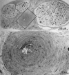
Epineurial arterioles were more frequently abnormal in nerves of diabetics than in nerves of controls. The arteriole enclosed by a rectangle in A and shown in greater detail in B, shows obliteration of the lumen by fat deposition, intimal proliferation, and thickening of the wall. (Reprinted with permission from Dyck PJ, Lais A, Karnes JL, et al. Fiber loss is primary and multifocal in sural nerves in diabetic polyneuropathy. Ann Neurol, 19:425–439, 1986.)
Figure 5
Figure 5
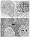
Transverse epoxy sections (p-phenylenediamine) of distal sural nerves from patients with diabetic lumbosacral radiculoplexus neuropathy illustrating the dramatic focal fiber loss characteristic of the disorder (A, fascicle on the left) and the abortive microfascicular nerve regeneration (B, as identified by the arrow). Note that the abortive regeneration (injury neuroma) is made up of multiple regenerating fascicles and that they are situated adjacent to a fascicle devoid of myelinated fibers. Most of the fibers in the right fascicle in the lower panel are actively degenerating. As discussed in the text, these changes are indicative of ischemic injury that we attribute to a microscopic vasculitis. (Reprinted with permission from Dyck PJB, Norell JE, Dyck PJ. Microvasculitis and ischemia in diabetic lumbosacral radiculoplexus neuropathy. Neurology, 53:2113–2121, 1999.)
Figure 6
Figure 6
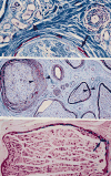
Transverse paraffin sections of sural nerves showing changes seen in DLRPN and LRPN. Upper panel, Masson’s trichrome stain, showing inflammation in the wall of an epineurial microvessel (right upper), probably fibrinoid degeneration of the perineurium (long arrow), and a region of neovascularization (arrowhead). Middle panel, Luxol fast blue-periodic acid Schiff stain, showing several fascicles surrounded by normal thickness perineurium (right middle, between the arrowheads) and one fascicle with extremely thick perineurium (left middle, between the arrowheads). We attribute the latter finding to scarring and repair after ischemic injury (note all fascicles are devoid of myelinated fibers). Lower panel, Turnbull blue stain, showing accumulation of hemosiderin (iron stains bright blue, arrow) deposited along the inner aspects of the perineurium. All of these pathological features are frequently seen together and are best explained by ischemic injury. (Reprinted with permission from Dyck PJB, Windebank AJ. Diabetic and nondiabetic lumbosacral radiculoplexus neuropathies: New insights into pathophysiology and treatment. Muscle Nerve, 25:477–491, 2002.)
Figure 7
Figure 7
Serial skip paraffin sections of a microvessel above (upper panel), at (middle panel), and below (lower panel) a region of microvasculitis in the sural nerve of a patient with diabetic lumbosacral radiculoplexus neuropathy. The sections on the left column are stained with hematoxylin and eosin, the sections in the middle column are reacted to antihuman smooth muscle actin (Dako), and the sections on the right column are reacted to leukocytes (CD 45). The smooth muscle of the tunica media in the region of microvasculitis (middle panel) is separated by mononuclear cells, fragmented and decreased in amount. The changes are those of a focal microvasculitis. (Reprinted with permission from Sinnreich M, Taylor BV, Dyck PJB. Diabetic neuropathies classification, clinical features and pathophysiological basis. The Neurologist, 11(2):63–79, 2005.)

References

    1. Leyden E. Beitrag zur Klinik des diabetes mellitus. Wien Med Wochenschr. 1893;43:926.
    1. Dyck PJB, Dyck PJ. Paresthesia, pain and weakness in hands of diabetic patients is attributable to mononeuropathies or radiculopathy, not polyneuropathy: The Rochester (RDNS) and Pancreas Renal Transplant (MC-PRT) Studies. Neurology. 1998;50:A333.
    1. Tracy JA, Dyck PJB, Harper CM, et al. Hand symptomatology in diabetes usually due to mononeuropathy, not polyneuropathy. Ann Neurol. 2005;58(Suppl 9):S36.
    1. Dyck PJ, O’Brien PC, Litchy WJ, et al. Monotonicity of nerve tests in diabetes. Subclinical nerve dysfunction precedes diagnosis of polyneuropathy. Diabetes Care. 2005;28(9):2192–200. - PubMed
    1. Gregersen G. Diabetic neuropathy: Influence of age, sex metabolic control, and duration of diabetes on motor conduction velocity. Neurology. 1967;17:972–80. - PubMed

Publication types

MeSH terms