

Review
doi: 10.1016/j.sbi.2007.11.003.
Epub 2008 Jan 14.
Junction-forming aquaporins
Affiliations
- PMID: 18194855
- PMCID: PMC2396442
- DOI: 10.1016/j.sbi.2007.11.003
Item in Clipboard
Review
Junction-forming aquaporins
Curr Opin Struct Biol.
2008 Apr.
Abstract
Aquaporins (AQPs) are a family of ubiquitous membrane channels that conduct water and solutes across membranes. This review focuses on AQP0 and AQP4, which in addition to forming water channels also appear to play a role in cell adhesion. We discuss the recently determined structures of the membrane junctions mediated by these two AQPs, the mechanisms that regulate junction formation, and evidence that supports a role for AQP0 and AQP4 in cell adhesion.
Figures

Cleavage of the cytoplasmic termini of AQP0 enhances the adhesive properties of its extracellular surface. (a) Proteoliposomes containing full-length AQP0 isolated from the lens cortex are uniformly distributed. (b) Treatment of the proteoliposomes shown in (a) with chymotrypsin causes the vesicles to cluster. (c) Proteoliposomes containing a mixture of full-length and truncated AQP0 isolated from the lens core cluster in a similar way as the proteoliposomes containing full-length AQP0 that were treated with chymotrypsin. (d) Reconstitution of full-length AQP0 isolated from the lens cortex produces single-layered 2D crystals. The inset shows a 4Å projection map of an AQP0 tetramer in the single-layered crystals. (e) Reconstitution of a mixture of full-length and truncated AQP0 isolated from the lens core produces double-layered 2D crystals. The arrowheads indicate the edges of the two crystal layers. The inset shows a 4Å projection map of two stacked AQP0 tetramers in the double-layered crystals. The diagonal black lines indicate the two mirror axes that result from the opposite orientations of the two interacting crystal layers. The scale bars in panels (a) to (e) correspond to 1 μm. The insets in (d) and (e) show complete unit cells with lattice constants of a = b = 65.5Å. Adapted from [20••], with permission from Elsevier.
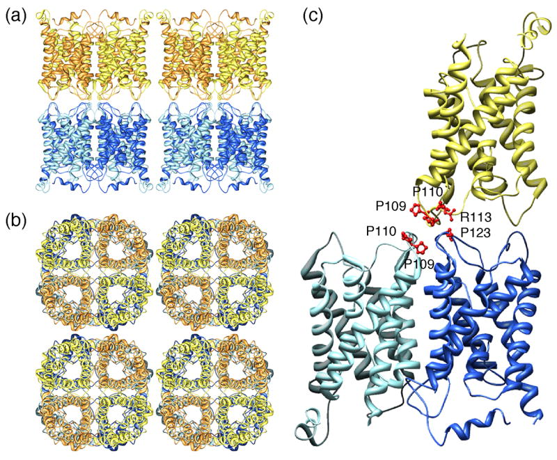
Double-layered 2D crystals formed by AQP0 (pdb accession code: 2B6O) [22••]. (a) View parallel and (b) view perpendicular to the membrane plane showing that the AQP0 tetramers in the two membrane layers are exactly in register. (c) Interactions between AQP0 tetramers formed by extracellular loop C. The proline-proline motif (Pro109 and Pro110) in loop C in the AQP0 monomer in the top layer (yellow) makes interaction with the proline-proline motif in an AQP0 monomer in the bottom layer (light blue). In addition, Arg113 in loop C in the AQP0 monomer in the top layer (yellow) makes interaction with Pro123 in loop C in another AQP0 monomer in the bottom layer (dark blue).
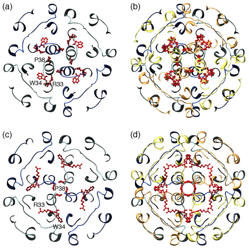
Two different conformations of AQP0’s extracellular loop A and the resulting interactions in paired tetramers. (a) Conformation of loop A in the 2.2Å X-ray structure of full-length AQP0 (pdb accession code: 1YMG) [27•]. Pro38 points away from the center of the tetramer, Trp34 lies above the pore and projects outward, blocking the approach of a second tetramer, and Arg33 is positioned in between two monomers. (b) The conformation of loop A in the paired tetramers seen in loosely packed 3D crystals of full-length AQP0 is the same as in (a), since the 2.2Å X-ray structure of full-length AQP0 was used to obtain this low resolution molecular replacement solution, but the two tetramers are rotated with respect to each other by 24° (pdb accession code: 2C32) [29•]. (c) Conformation of loop A in the double-layered 2D crystals (pdb accession code: 2B6O) [22••]. Pro38 forms a rosette-like structure at the center of the tetramer, and the side chains of Arg33 and Trp34 have swapped positions (compare with panel a). (d) In the completed junction, residues Pro38, Arg33 and Trp34 interact with the corresponding residues from the opposing tetramer.
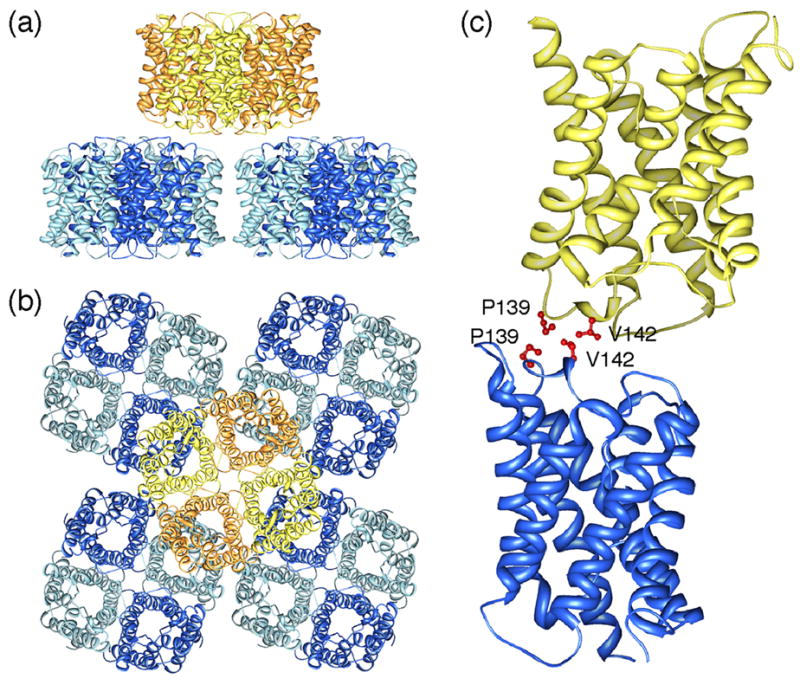
Double-layered 2D crystals formed by AQP4 (pdb accession code: 2D57) [39••]. (a) View parallel and (b) view perpendicular to the membrane plane showing that an AQP4 tetramer in one membrane layer makes interactions with four AQP4 tetramers in the other membrane layer. (c) Interactions between tetramers in the two layers are formed by residues Pro139 and Val142 in AQP4’s extracellular loop C.
References
-
- Tsukaguchi H, Shayakul C, Berger UV, Mackenzie B, Devidas S, Guggino WB, van Hoek AN, Hediger MA. Molecular characterization of a broad selectivity neutral solute channel. J Biol Chem. 1998;273:24737–24743. - PubMed
-
- Yasui M, Hazama A, Kwon TH, Nielsen S, Guggino WB, Agre P. Rapid gating and anion permeability of an intracellular aquaporin. Nature. 1999;402:184–187. - PubMed
-
- Maurel C. Plant aquaporins: novel functions and regulation properties. FEBS Lett. 2007;581:2227–2236. - PubMed
Publication types
MeSH terms
Substances
Grants and funding
LinkOut - more resources
Full Text Sources
