. 2008 Jan 29;105(4):1134-9.
doi: 10.1073/pnas.0711168105. Epub 2008 Jan 14.

HSP90 regulates cell survival via inositol hexakisphosphate kinase-2

Affiliations

HSP90 regulates cell survival via inositol hexakisphosphate kinase-2

Anutosh Chakraborty et al. Proc Natl Acad Sci U S A. .

Abstract

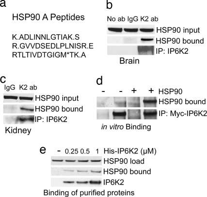
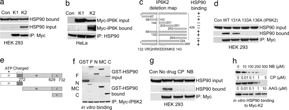
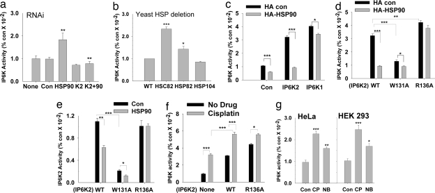
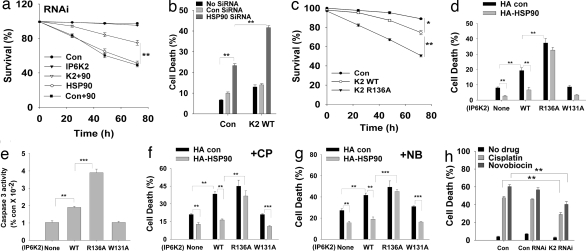
Heat-shock proteins (HSPs) are abundant, inducible proteins best known for their ability to maintain the conformation of proteins and to refold damaged proteins. Some HSPs, especially HSP90, can be antiapoptotic and the targets of anticancer drugs. Inositol hexakisphosphate kinase-2 (IP6K2), one of a family of enzymes generating the inositol pyrophosphate IP7 [diphosphoinositol pentakisphosphate (5-PP-IP5)], mediates apoptosis. Increased IP6K2 activity sensitizes cancer cells to stressors, whereas its depletion blocks cell death. We now show that HSP90 physiologically binds IP6K2 and inhibits its catalytic activity. Drugs and selective mutations that abolish HSP90-IP6K2 binding elicit activation of IP6K2, leading to cell death. Thus, the prosurvival actions of HSP90 reflect the inhibition of IP6K2, suggesting that selectively blocking this interaction could provide effective and safer modes of chemotherapy.


Conflict of interest statement

The authors declare no conflict of interest.

Figures

Fig. 1.
Fig. 1.
IP6K2 binds HSP90. (a) HSP90A peptides identified by LC/MS/MS analysis. (b) Co-IP of endogenous HSP90 by endogenous IP6K2 in mouse brain. Coprecipitated HSP90 was detected by Western blotting using a monoclonal antibody. (c) HSP90 coprecipitated with IP6K2 in mouse kidney. Immunoprecipitation was done with IgG- or IP6K2-specific antibody as in brain extract. HSP90 in the immunoprecipitated sample was detected by Western blotting. (d) Direct binding of immunoprecipitated Myc-IP6K2 with exogenously added HSP90 purified from HeLa cells. Myc-vector-transfected HEK293 cells were used as negative controls. (e) Direct binding of purified recombinant His-IP6K2 with exogenously added HSP90 purified from HeLa cells.
Fig. 2.
Fig. 2.
HSP90 binds to a specific motif in IP6K2 by its C terminus. (a) Co-IP of endogenous HSP90 by overexpressed Myc-IP6K2 from HEK293 cells. Protein (1 mg) from each cell lysate was immunoprecipitated by anti-Myc antibody and was immunoblotted with monoclonal HSP90 antibody. Lane 1 (Con) shows the Myc-vector control. (b) Co-IP of Myc-IP6K2, not IP6K1, by endogenous HSP90 from HeLa cells. Immunoprecipitation of endogenous HSP90 by monoclonal antibody coprecipitates IP6K2, not IP6K1, as confirmed by blotting with α-Myc antibody. (c) Deletion mapping of IP6K2 to identify HSP90-binding motif. (d) Endogenous HSP90 does not coprecipitate with mutants of IP6K2 in the putative HSP90-binding region. R133A and R136A mutants do not bind, whereas W131A has little effect on binding. (e) Mapping of HSP90 to identify the IP6K2-binding region. Fragments 1–272 [N terminus (N)], 273–732 [middle and C termini (MC)], and 629–732 [C terminus (C)] were generated and cloned into pGEX vector and purified from bacteria. (f) Determination of binding region of HSP90 to IP6K2 by in vitro binding. After incubation of the proteins, the beads were washed, and bound HSP90 was analyzed by blotting with anti-HSP90 monoclonal antibody. (g) IP6K2–HSP90 interaction in cells is disrupted by CP and NB treatment. Overnight drug treatment was followed by immunoprecipitation of Myc-IP6K2 and detection of coprecipitated HSP90 by Western blotting. (h) Binding in vitro of HSP90–IP6K2 is disrupted by CP and NB, but not by AAG. HSP90 bound to Myc-IP6K2 was analyzed by blotting with anti-HSP90 monoclonal antibody.
Fig. 3.
Fig. 3.
HSP90 inhibits IP6K2 catalytic activity. (a) IP6K2 activity in vivo is increased in the absence of endogenous HSP90. HSP90 and IP6K2 (either alone or together) were depleted by using siRNA in [3H]inositol-labeled HEK293 cells and inositol phosphates separated by HPLC. (b) IP6K activity in WT and HSP mutant S. cerevisiae in vivo. IP6 and IP7 were monitored after [3H]inositol labeling of intact cells. (c) HSP90 overexpression leads to decreased IP6K2 activity in vivo. HEK293 cells overexpressing Myc-IP6K1/Myc-IP6K2 either alone or with HA-HSP90 were labeled with [3H]inositol, and inositol phosphates were isolated by HPLC. (d) HSP90 inhibition of IP6K2 in vivo requires binding to IP6K2. IP7 labeled by [3H]inositol was assessed in HEK293 cells cotransfected with Myc-IP6K2 (WT or the mutants) and HA-HSP90. (e) IP6K activity in vitro of WT and mutant IP6K2 in the absence and presence of purified HSP90. HSP90 fails to inhibit IP6K activity of the R136A mutant that does not bind HSP90. IP6K2-W131A is catalytically inactive. (f) CP enhancement of IP7 generation in vivo is influenced by IP6K2–HSP90 binding. IP6K activity of [3H]inositol-labeled HEK293 cells was measured after 30 μM CP treatment overnight. (g) IP6K activity is increased after CP and NB treatment of HEK293 and HeLa cells. After drug treatment, extracts were prepared, and 25 μg of total protein was assayed for IP6K activity.
Fig. 4.
Fig. 4.
HSP90–IP6K2 interaction regulates drug-induced cell death. (a) Decreased cell survival elicited by HSP90 depletion (filled inverted triangles) is reversed by codepletion of IP6K2 (filled squares). Cotansfection of control siRNA with HSP90 siRNA has no effect (open squares). MTT assay was done after each time period of various siRNA treatments of HEK293 cells. (b) Cell death elicited by HSP90 depletion is further enhanced by IP6K2 overexpression. Cells were either transfected with HSP90 siRNA or cotransfected with Myc-IP6K2 for 48 h, and cell death was monitored by apoptotic nuclei-detection assay. (c) Cell survival (MTT) assay of WT and IP6K2-R136A-transfected HEK293 cells. Cells were transfected with various Myc-IP6K2 constructs; after each interval, the percentage survival was calculated. IP6K2-R136A mutant (filled inverted triangles) causes a significant decrease in cell survival than IP6K2-WT (open circles). (d) Increased cell death elicited by overexpression of IP6K2-R136A that does not bind to HSP90. Cells were transfected by different constructs for 72 h. Cell death caused by IP6K2-WT is reversed by HSP90 overexpression, whereas there is no effect of HSP90 on IP6K2-R136A. The catalytically impaired IP6K2-W131A does not induce cell death. (e) Caspase-3 activity is increased in cells transfected with IP6K2-R136A, which does not bind HSP90. Activity was measured after 72 h of transfection. The magnitude of increase was determined by considering OD405 of control samples as unity. (f) Death of cells overexpressing IP6K2-WT after CP treatment is reversed by HSP90 in cells overexpressing IP6K2-WT, but not IP6K2-R136A, which cannot bind HSP90. Cells overexpressing IP6K2-W131A show diminished cell death relative to WT, which is further reduced by HSP90 coexpression. (g) Cell death assay as described for panel f, after NB treatment. (h) CP- and NB-induced cell death in HEK293 cells requires IP6K2. Endogenous IP6K2 was depleted by using siRNA. CP and NB were added after 36 h of transfection for 36 and 24 h, respectively. Cell death in drug-treated cells is diminished by IP6K2 depletion.

Comment in

References

    1. Berridge MJ, Lipp P, Bootman MD. Signal transduction: The calcium entry pas de deux. Science. 2000;287:1604–1605. - PubMed
    1. Irvine RF, Schell MJ. Back in the water: The return of the inositol phosphates. Nat Rev Mol Cell Biol. 2001;2:327–338. - PubMed
    1. Menniti FS, Miller RN, Putney JW, Jr, Shears SB. Turnover of inositol polyphosphate pyrophosphates in pancreatoma cells. J Biol Chem. 1993;268:3850–3856. - PubMed
    1. Stephens L, et al. The detection, purification, structural characterization, and metabolism of diphosphoinositol pentakisphosphate(s) and bisdiphosphoinositol tetrakisphosphate(s). J Biol Chem. 1993;268:4009–4015. - PubMed
    1. Saiardi A, Erdjument-Bromage H, Snowman AM, Tempst P, Snyder SH. Synthesis of diphosphoinositol pentakisphosphate by a newly identified family of higher inositol polyphosphate kinases. Curr Biol. 1999;9:1323–1326. - PubMed

Publication types

MeSH terms

LinkOut - more resources