. 2008 Jan 22;105(3):895-900.
doi: 10.1073/pnas.0707284105. Epub 2008 Jan 14.

Universality and diversity of folding mechanics for three-helix bundle proteins

Affiliations

Universality and diversity of folding mechanics for three-helix bundle proteins

Jae Shick Yang et al. Proc Natl Acad Sci U S A. .

Abstract

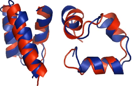
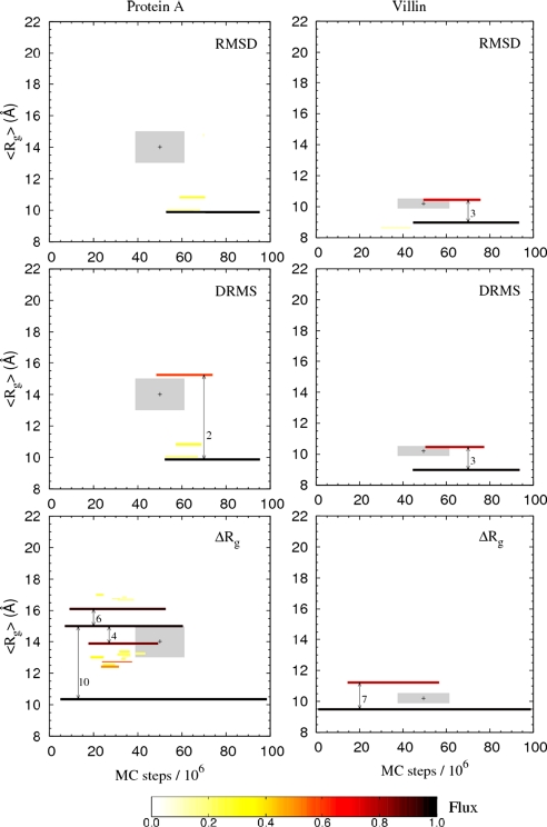
In this study we evaluate, at full atomic detail, the folding processes of two small helical proteins, the B domain of protein A and the Villin headpiece. Folding kinetics are studied by performing a large number of ab initio Monte Carlo folding simulations using a single transferable all-atom potential. Using these trajectories, we examine the relaxation behavior, secondary structure formation, and transition-state ensembles (TSEs) of the two proteins and compare our results with experimental data and previous computational studies. To obtain a detailed structural information on the folding dynamics viewed as an ensemble process, we perform a clustering analysis procedure based on graph theory. Moreover, rigorous p(fold) analysis is used to obtain representative samples of the TSEs and a good quantitative agreement between experimental and simulated Phi values is obtained for protein A. Phi values for Villin also are obtained and left as predictions to be tested by future experiments. Our analysis shows that the two-helix hairpin is a common partially stable structural motif that gets formed before entering the TSE in the studied proteins. These results together with our earlier study of Engrailed Homeodomain and recent experimental studies provide a comprehensive, atomic-level picture of folding mechanics of three-helix bundle proteins.


Conflict of interest statement

The authors declare no conflict of interest.

Figures

Fig. 1.
Fig. 1.
Comparison between the native structures (in blue) and superimposed minimum-energy top-k structures (in red) obtained through a clustering procedure for protein A (Left) and Villin (Right), as described in the text. The rmsd values between top-k and experimental structures are 2.7 and 2.1 Å for protein A and Villin, respectively. Structures were created by using PyMOL (50).
Fig. 2.
Fig. 2.
Relaxation behavior of the average radius of gyration, Rg (Upper) and average fraction helicity (Lower) of each individual helix, obtained at T ≈ 300 K. Helicity is determined with the criterion of Kabsch and Sander (51). The Rg relaxation data are fitted (red curves, Upper) to a double-exponential function, f(t) = a1 exp(−tfast) + a2 exp(−tslow) + b, using a Levenberg–Marquardt fit procedure with a1, a2, τfast, τslow, and b as free parameters.
Fig. 3.
Fig. 3.
Results of the structural kinetic cluster analysis for protein A and Villin. Each cluster is represented by a line, from t = MFPT to t = MLET, and color-coded by its flux, F, as indicated by the color scale. Only clusters with F > 0.1 are shown. The vertical location of each cluster is determined by the average radius of gyration, <Rg>, where < > is the cluster average. The location of the two TSEs for protein A and Villin are indicated by shaded areas centered (+) around the average Rg and time t, with averages taken over the two ensembles; the sizes of the two shaded areas reflect 1σ deviations in both the Rg and t directions. The rmsd structural graphs are obtained by using the cutoffs dc = 1.1 Å and 1.5 Å for protein A and Villin, respectively, whereas for drms we use dc = 0.9 Å and 1.2 Å, respectively. With these choices of dc, the rmsd and drms GCs contain ≈35% of all conformations, a reasonable number given that the trajectory time is ≈2τslow. For ΔRg, all reasonable choices of dc give larger GCs than for rmsd and drms, indicating that it contains not only native-like structures (see text). The results for ΔRg are not very sensitive to the specific choice of dc. Results are shown obtained for dc = 0.0080 Å and 0.0040 Å, respectively.
Fig. 4.
Fig. 4.
Comparison between simulated and experimental (32) Φ values. At positions where more than one experimental Φ value is reported, the average value is used, and their individual experimental Φ values on different types of mutations are shown in SI Fig. 9. Error bars denote the standard deviation, σ.

References

    1. Alm E, Baker D. Curr Opin Struct Biol. 1999;9:189–196. - PubMed
    1. Gianni S, Guydosh NR, Khan F, Caldas TD, Mayor U, White GW, DeMarco ML, Daggett V, Fersht AR. Proc Natl Acad Sci USA. 2003;100:13286–13291. - PMC - PubMed
    1. Laurents DV, Baldwin RL. Biophys J. 1998;75:428–434. - PMC - PubMed
    1. Pande VS. Proc Natl Acad Sci USA. 2003;100:3555–3556. - PMC - PubMed
    1. Lo Conte L, Ailey B, Hubbard TJ, Brenner SE, Murzin AG, Chothia C. Nucleic Acids Res. 2000;28:257–259. - PMC - PubMed

Publication types

LinkOut - more resources