. 2008 Jul;75(7):1143-53.
doi: 10.1002/mrd.20856.

Spatial alignment of the mouse blastocyst axis across the first cleavage plane is caused by mechanical constraint rather than developmental bias among blastomeres

Affiliations

Spatial alignment of the mouse blastocyst axis across the first cleavage plane is caused by mechanical constraint rather than developmental bias among blastomeres

Vernadeth B Alarcón et al. Mol Reprod Dev. 2008 Jul.

Abstract

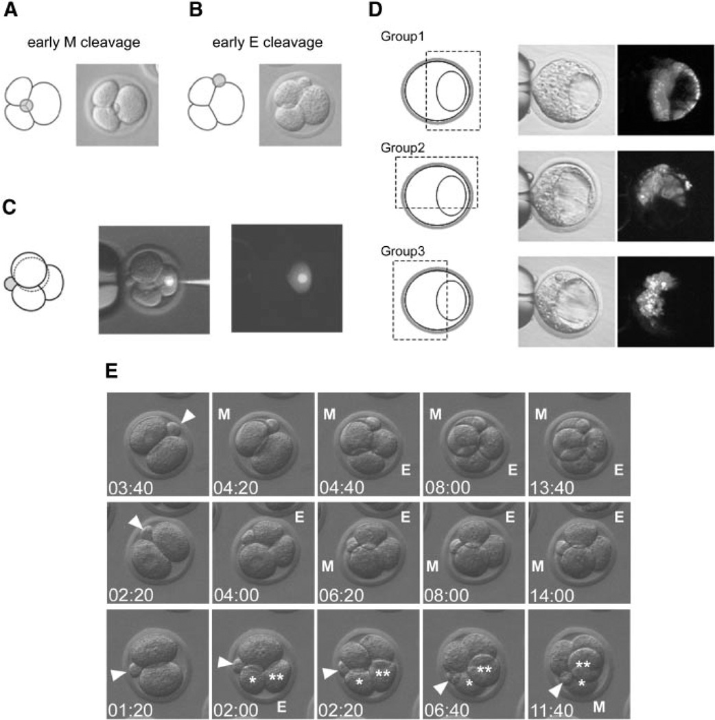
The embryonic-abembryonic (Em-Ab) axis of the mouse blastocyst has been found in several studies to align orthogonal to the first cleavage plane, raising the possibility that a developmental prepattern already exists at the two-cell stage. However, it is also possible that such alignment is not due to any developmental disparity between the two-cell stage blastomeres, but rather is caused by an extrinsic mechanical constraint that is conferred by an irregular shape of the zona pellucida (ZP). Here, we conducted a series of experiments to distinguish between these possibilities. We showed that the shape of the ZP at the two-cell stage varied among embryos, ranging from near spherical to ellipsoidal, and that the ZP shape did not change until the blastocyst stage. In those embryos with an ellipsoidal ZP, the Em-Ab axis tended to lie orthogonal to the first cleavage plane, while in those embryos with a near spherical ZP, there was no such relationship. The clonal boundary between the descendants of the two-cell stage blastomeres tended to lie orthogonal to the Em-Ab axis when the rotation of the embryo within the ZP was experimentally prevented, while the control embryos did not exhibit such tendency. These results support the possibility that an apparent correlation between the first cleavage plane and the blastocyst axis can be generated by the mechanical constraint from the ZP but not by a developmental prepattern. Moreover, recent reports indicate that the vegetal blastomere of the four-cell stage embryo that had undergone a specific type of second cleavages is destined to contribute to the abembryonic side of the blastocyst. However, our present study shows that in spite of such specific second cleavages, the vegetal blastomere did not preferentially give rise to the abembryonic side. This result implicates that the lineage of the four-cell stage blastomere is not restricted even when embryos undergo a specific type of second cleavages.


Figures

Fig. 1
Fig. 1
A: A blastocyst develops within the zona pellucida (ZP), and is composed of two distinct cell populations, that is, inner cell mass (ICM) and trophectoderm (TE). The embryonic–abembryonic axis of the blastocyst is defined based on the location of ICM relative to the blastocyst cavity. B: A schematic diagram depicting how the mechanical constraint model can explain some of the observations which the prepattern model is based on (see text for details). C: The orientations of the second cleavages are categorized into meridional (M) and equatorial (E) with respect to the animal–vegetal axis (see text for details).
Fig. 2
Fig. 2
Persistence of the ellipsoidal ZP shape throughout early development. A: The internal diameters of ZP are measured in three ways relative to the first cleavage plane and the position of the second polar body, as depicted in the figures. B: Mean, standard deviation (SD), and range of the ratios between the internal diameters of the ZP in the embryos examined (n = 46). C: Examples of embryos that were monitored by time-lapse videomicroscopy (n = 50). Images at the two-cell, four-cell, eight-cell, and morula stages are shown. The inner surface of the ZP for each embryo is highlighted with a white broken line. Note that the shape of the ZP persists regardless of the developmental stages of the embryos.
Fig. 3
Fig. 3
Relationship among the ZP shape, the first cleavage plane, and the Em–Ab axis. A: The shape of ZP is evaluated at the two-cell stage based on the ratio between the two internal diameters: one (a) is perpendicular to and the other (b) passes through the first cleavage plane. B: The angle (θ) between the first cleavage plane and the blastocyst cavity floor is measured at the early blastocyst stage. C,C’,D,D’: Two embryos are shown as examples of those (n = 41) that were monitored by time-lapse videomicroscopy. E: A scatter plot of the ZP shape (b/a) and the angle (θ). The data are summarized in Table 1.
Fig. 4
Fig. 4
The clonal boundary between the two-cell stage blastomeres tends to lie orthogonal to the Em–Ab axis when the rotation of the embryo within the ZP is prevented. A: A schematic diagram describing how to fill the perivitelline space of the DiI-labeled embryos with alginate gel (see text for details). B: Two examples of early blastocysts that were derived from alginate-filled, two-cell stage embryos. Left panels are bright field images, middle panels are fluorescence images showing the distribution of DiI-labeled cells, and right panels are overlay of the two images with projected lines for the blastocyst cavity floor (black broken line) and the boundary between the labeled and nonlabeled cells (white broken line). C: A graph showing the occurrence of angles (θ in A) in the alginate-filled embryos (gray bars; n = 47) and control embryos (white bars; n = 44). The angles are categorized into three ranges. D: The same data as presented in (C), with the angles categorized into six ranges. E: Examples of control (top two rows) and alginate-filled (bottom two rows) embryos to show that alginate gel diminishes the movement of embryos within the ZP. The embryo together with the attached polar body (arrowhead) tends to rotate within the ZP during early cleavages in control cases, while such rotation is markedly less in alginate-filled cases. From left to right, columns represent the two-cell, the early eight-cell, the late eight-cell, and the beginning of the blastocyst stages.
Fig. 5
Fig. 5
Lineage tracing of the vegetal blastomere of the ME-type of four-cell stage embryos. A: A three-cell stage embryo in which the early second cleavage had occurred in a meridional (M) orientation. B: A three-cell stage embryo in which the early second cleavage had occurred in an equatorial (E) orientation. C: The DiI-labeling of the vegetal blastomere in the four-cell stage embryo that had undergone the ME-type of second cleavages. D: The distribution of the descendants of the labeled ME-vegetal blastomere at the early blastocyst stage. The distribution patterns are categorized into three groups (see text for details) and are summarized in Table 2. E: Examples of embryos that were monitored by time-lapse video-microscopy from the two-cell to the late four-cell stages to demonstrate the position of the second polar body relative to the second cleavage planes. In each frame, time is given in hours:minutes after the start of the time-lapse recording. Embryo in the top row is undergoing ME-type of second cleavages, while embryo in the middle row is undergoing EM-type of second cleavages. Embryo in the bottom row shows the early equatorial cleavage producing blastomeres (*,**) that shifted position so that they appear as having undergone the meridional cleavage.

References

    1. Alarcón VB, Marikawa Y. Deviation of the blastocyst axis from the first cleavage plane does not affect the quality of mouse postimplantation development. Biol Reprod. 2003;69:1208–1212. - PubMed
    1. Alarcón VB, Marikawa Y. Unbiased contribution of the first two blastomeres to mouse blastocyst development. Mol Reprod Dev. 2005;72:354–361. - PubMed
    1. Beddington RSP, Robertson EJ. Axis development and early asymmetry in mammals. Cell. 1999;96:195–209. - PubMed
    1. Chroscicka A, Komorowski S, Maleszewski M. Both blastomeres of themouse 2-cell embryo contribute to the embryonic portion of the blastocyst. Mol Reprod Dev. 2004;68:308–312. - PubMed
    1. Committee to Revise the Guide, Institute of Laboratory Animal Resources Council, Commission on Life Sciences, National Research Council. Guide for the Care and Use of Laboratory Animals. 7th edition. Washington, DC: National Academy Press; 1996.

Publication types

LinkOut - more resources