. 2008 Feb 12;47(6):1631-9.
doi: 10.1021/bi7016359. Epub 2008 Jan 16.

Self-association of human PCSK9 correlates with its LDLR-degrading activity

Affiliations

Self-association of human PCSK9 correlates with its LDLR-degrading activity

Daping Fan et al. Biochemistry. .

Abstract

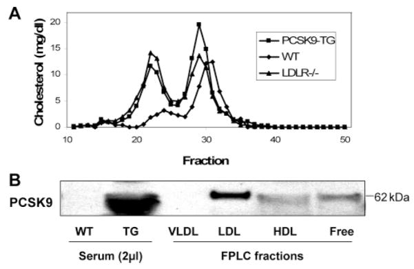
Genetic studies have demonstrated an important role for proprotein convertase subtilisin/kexin type 9 (PCSK9) as a determinant of plasma cholesterol levels. However, the underlying molecular mechanism is not completely understood. To this end, we have generated a mammalian cell expression system for human PCSK9 and its mutants and produced transgenic mice expressing human PCSK9. HEK293T cells transfected with the human PCSK9 DNA construct expressed and secreted PCSK9 and displayed decreased LDLR levels; functional PCSK9 protein was purified from the conditioned medium. In vitro studies showed that PCSK9 self-associated in a concentration-, temperature-, and pH-dependent manner. A mixture of PCSK9 monomers, dimers, and trimers displayed an enhanced LDLR degrading activity compared to monomeric PCSK9. A gain-of-function mutant, D374Y, displayed greatly increased self-association compared to wild-type PCSK9. Moreover, we demonstrated that the catalytic domain of PCSK9 is responsible for the self-association. Self-association of PCSK9 was enhanced by incubation with mouse apoE-/- VLDL and inhibited by incubation with both human and mouse HDL. When PCSK9 protein was incubated with total serum, it partially associated with LDL and HDL but not with VLDL. In transgenic mice, PCSK9 also associated with LDL and HDL but not with VLDL. We conclude that self-association is an intrinsic property of PCSK9, correlated to its LDLR-degrading activity and affected by plasma lipoproteins. These results provide a basis for developing strategies to manipulate PCSK9 activity in the circulation for the treatment of hypercholesterolemia.


Figures

Figure 1
Figure 1
Expression of PCSK9 and its functional assessment. A. HEK293T cells were transfected with pcDNA-hPCSK9. Cell lysate and conditioned medium were subjected to 4–12% SDS-PAGE and western blot analysis using anti-HisTag Ab for hPCSK9. P, pro-PCSK9; M, mature PCSK9. B. Cell lysates from pcDNA-PCSK9 transfected (+PCSK9) or non-transfected (−PCSK9) HEK293T cells were blotted for PCSK9, LDLR, and β-actin. C. Purified PCSK9 was added to HEK293T cell membrane preparation at 6 μg/mL. After incubation at 37 °C for 16 h, the membrane preparation was subjected to western blot analyses for LDLR and transferrin receptor. D. Purified PCSK9 was added to HEK293T cell culture medium at 6 μg/mL. After incubation at 37 °C for 16 h, whole cell extracts were subjected to western blot analysis of LDLR and β-actin. Three experiments showed LDLR was reduced by 80% on average by incubation with PCSK9.
Figure 2
Figure 2
Self-association of PCSK9 and its impact on LDLR degrading activity. A. Purified hPCSK9 was incubated at indicated concentration and temperature for 24 h before subjected to western blot analysis. De, a degraded fragment. B. Before added to HEK293T cells, purified PCSK9 protein at indicated concentration was incubated in DMEM without FBS for 24 h at 4 °C or 37 °C. Then the media were added to HEK293T cells and the cells were incubated at 37 °C for an additional 4 h. The whole cell lysates were subjected to western blot analyses of LDLR and β-actin. Normalized ratios of LDLR to β-actin were measured by densitometry with Quantity One software in three separate experiments and the average numbers are shown.
Figure 3
Figure 3
Self-association of PCSK9 mutants. A. HEK293T cells were transfected with wild-type (WT) PCSK9, a gain-of-function mutant D374Y and a loss-of-function mutant C679X; 24 h after transfection, the medium and cell lysate were subjected to western blot analysis of PCSK9 (rabbit anti-PCSK9 antibody) and LDLR. B. Purified Wild-type PCSK9 and D374Y mutant were incubated at 1 μg/mL, 37 °C and pH 5.2 or pH 7.0 for 24 h, and then subjected to western blot analysis using rabbit anti-PCSK9 antibody. C. Purified WT PCSK9 and its domain-deletion mutants were incubated at 4 °C or 37 °C for 24 h at 5 μg/mL concentration and pH 7.0 and then were subjected to western blot analysis. D. Cross-linking of WT PCSK9 and its domain-deletion mutants at a concentration of 0.5 μg/mL (see Experimental Procedures). In both panel C and panel D, Lanes 1 and 4: full-length PCSK9; Lanes 2 and 5: PCSK9-RCD; Lanes 3 and 6: PCSK9-RPSCA.
Figure 4
Figure 4
Self-association of differently tagged PCSK9. A. Purified PCSK9, either with HisTag or GST tag, formed SDS-stable dimers and trimers after incubation at 12 μg/mL concentration, pH 5.2 and 37 °C for 4 h. B. Purified His-tagged PCSK9 and GST-tagged PCSK9 were mixed and incubated at 37 °C for 10 min and then pulled down by GST-affinity beads, and the pulled-down protein was subjected to western blot using rabbit anti-PCSK9 antibody.
Figure 5
Figure 5
Uptake of PCSK9 multimers by cells. A. Purified PCSK9 was incubated in DMEM at 12 μg/mL and 37 °C for 24 h before being added to cultured HEK293T cells. The medium was then sampled at indicated time points for western blot analysis of PCSK9. B. Self-association of secreted PCSK9 in the culture medium of transfected HEK293T cells. After the cells were transfected in a 10-ch dish with PCSK9 for 24 h, half of the medium was transferred to an empty dish (no cells cultured) with protease inhibitor cocktail added. After further incubation at 37 °C for 24 h, the medium containing the cells (lane 1) and the cell-free medium (lane 2) were subjected to western blot. C. Intracellular presence of PCSK9 multimers. The cell-free medium containing PCSK9 was concentrated by Amicon Ultra centrifugal filter device to 1/10 of the original volume and added to cultured HEK293T cells along with 9 times the volume of 0.15 M KCl solution, and the cells were further cultured for 1 or 2 h. The cells were washed with 0.15 M KCl solution three times and lysed for western blot analysis. Lane 1: concentrated PCSK9-containing medium; lane 2: cell lysate at 1 h time point; lane 3: cell lysate at 2 h time point; lane 4: control cell lysate without addition of PCSK9-containing medium.
Figure 6
Figure 6
Autocrinal and paracrinal effects of secreted PCSK9. A. Transduction of HEK293T cells with PWPI-PCSK9 lentiviruses diminished cellular LDLR; non-transduced cells and cells transduced with PWPI control lentiviruses were included as controls. B. Confocal microscopy images of transduced HEK293T cells. The cells were transduced with indicated MOI of lentiviruses. Green fluorescence is from GFP, and LDLR was stained in red in a non-permeablized condition. Note: in the middle panel, the white arrow-pointed 3 cells were not transduced, LDLR staining is stronger than the transduced cells in the same image, but weaker than the non-transduced cells in the left image.
Figure 7
Figure 7
Lipoproteins affect self-association of PCSK9. Purified PCSK9 protein at 12 μg/mL was incubated at 37 °C for 24 h with different lipoproteins (30 μg protein/mL). At the end of incubation, the protein was subjected to western blot using anti 6 × His antibody for PCSK9. Lane 1: 4 °C control; lane 2: 37 °C control without addition of lipoprotein; lanes 3–9, with addition of human VLDL, human LDL, human HDL, wild-type mouse VLDL, apoE−/− mouse VLDL, wild-type mouse HDL, and apoE−/− mouse HDL, respectively.
Figure 8
Figure 8
Purified PCSK9 associates with mouse lipoproteins. Purified PCSK9 (50 μg/mL) was incubated with mouse serum at 37 °C for 30 min. The serum (100 μL) was then subjected to FPLC. Cholesterol concentration was measured for each FPLC fraction (A). On the basis of cholesterol content, fractions were pooled to present VLDL (fractions 14–18), LDL (fractions 19–27), HDL (fractions 28–33), and cholesterol free (fractions 34–50) part. Each part was concentrated to 100 μL; 20 μL of each part was subjected to western blot analysis using anti 6 x His antibody (B).
Figure 9
Figure 9
Association of PCSK9 with lipoproteins in PCSK9 transgenic mouse serum. A. A pooled serum sample from three human PCSK9 transgenic mice was subjected to FPLC separation of lipoprotein fractions. Cholesterol content of each fraction was shown. Fractions were pooled to present VLDL (fractions 14–17), LDL (fractions 18–25), HDL (fractions 26–34), and cholesterol free (fractions 35–50). B. The pooled FPLC parts were concentrated to 250 μL each, and 30 μL from each part was loaded to SDS-gel for western blot analysis for PCSK9 using rabbit anti-PCSK9 antibody, 2 μL wild-type mouse serum, and PCSK9 transgenic mouse serum were loaded as controls.

References

    1. Seidah NG, Benjannet S, Wickham L, Marcinkiewicz J, Jasmin SB, Stifani S, Basak A, Prat A, Chretien M. The secretory proprotein convertase neural apoptosis-regulated convertase 1 (NARC-1): liver regeneration and neuronal differentiation. Proc Natl Acad Sci USA. 2003;100:928–933. - PMC - PubMed
    1. Naureckiene S, Ma L, Sreekumar K, Purandare U, Lo CF, Huang Y, Chiang LW, Grenier JM, Ozenberger BA, Jacobsen JS, Kennedy JD, DiStefano PS, Wood A, Bingham B. Functional characterization of Narc 1, a novel proteinase related to proteinase K. Arch Biochem Biophys. 2003;420:55–67. - PubMed
    1. Steiner D. The proprotein convertases. Curr Opin Chem Biol. 1998;2:31–39. - PubMed
    1. Zhou A, Webb G, Zhu X, Steiner DF. Proteolytic processing in the secretory pathway. J Biol Chem. 1999;274:20745–20748. - PubMed
    1. Seidah NG, Chretien M. Proprotein and prohormone convertases: a family of subtilases generating diverse bioactive polypeptides. Brain Res. 1999;848:45–62. - PubMed

Publication types