Review
. 2008 Apr 12;363(1495):1349-62.
doi: 10.1098/rstb.2007.2252.

Molecular analysis of neural crest migration

Affiliations
Review

Molecular analysis of neural crest migration

Sei Kuriyama et al. Philos Trans R Soc Lond B Biol Sci. .

Abstract

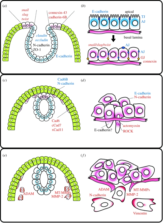
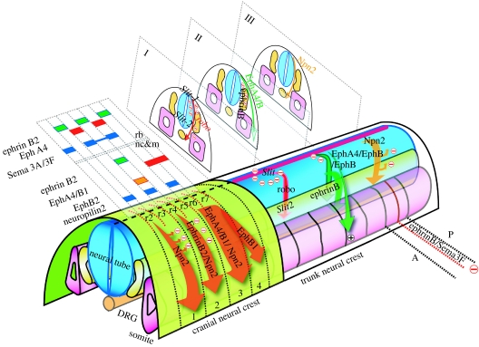
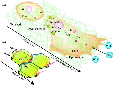
The neural crest (NC) cells have been called the 'explorers of the embryos' because they migrate all over the embryo where they differentiate into a variety of diverse kinds of cells. In this work, we analyse the role of different molecules controlling the migration of NC cells. First, we describe the strong similarity between the process of NC migration and metastasis in tumour cells. The epithelial-mesenchymal transition process that both kinds of cells undergo is controlled by the same molecular machinery, including cadherins, connexins, Snail and Twist genes and matrix metalloproteases. Second, we analysed the molecular signals that control the patterned migration of the cephalic and trunk NC cells. Most of the factors described so far, such as Eph/ephrins, semaphorins/neuropilins and Slit/Robo, are negative signals that prohibit the migration of NC cells into target areas of the embryo. Finally, we analyse how the direction of migration is controlled by regulation of cell polarity and how the planar cell polarity or non-canonical Wnt signalling is involved in this process.


Figures

Figure 1
Figure 1
NC migrations versus cancer metastasis. (a,b) Loss of polarity; (c,d) changes in cell adherence and cytoskeleton; (e,f) epithelial mesenchymal transition. (a,c,e) Different steps during NC migration. (b,d,f) Different steps of cell migration in cancer cells. Red letters represent factors upregulated and blue letters represent factors downregulated (purple cells, NC or metastatic cells; green cells, epidermis; blue cells, neural tube; AJ, adherence junction; TJ, tight junction; GJ, gap junction; see text for details).
Figure 2
Figure 2
Migration of cranial and trunk NC. Different factors that control the migration in the cranial and trunk NC are shown. Arrows represents streams of migrating NC. Cranial NC: migration in the head is controlled by Eph/ephrins and semaphorins/neuropilins. The expression of the different factors is shown in the rhombomeres (rb) and in the NC and mesoderm (nc&m). Trunk NC: migration in the trunk: I, ventromedial migration controlled by Slit/Robo; II, dorsolateral migration controlled by Eph/ephrins; III, ventromedial migration controlled by semaphorins/neuropilins. In addition, the anterior–posterior (A–P) patterning of NC migration is controlled by Eph/ephrins and semaphorins/neuropilins. See text for details.
Figure 3
Figure 3
Polarization of migrating NC in vivo. Differential interference contrast/time-lapse analysis of trunk NC migration in zebrafish embryos. Frames were taken every 3 min. In (a) neural tube (NT) and somites (S) are indicated (arrow, migrating NC; arrowheads, cell protrusions). (a) t=0 min, (b) t=3 min, (c) t=6 min, (d) t=9 min, (e) t=12 min and (f) t=15 min.
Figure 4
Figure 4
Polarization of migrating NC in vitro. Scanning electron micrograph of a Xenopus NC cultured in fibronectin. The polarity of the cells can be easily distinguished, with large lamellipodia and several fillopodia at the front of the cells and a retracting protrusion at the back. Arrow indicates the direction of migration.
Figure 5
Figure 5
PCP control polarity of migrating NC cells. (a) Tissue polarity in Drosophila. Several PCP factors such as Fz (red line), Dsh and Stbm (blue line) are localized in a polarized manner, leading to the polarization of the cytoskeleton (red mesh represent actin) and the formation of the bristle in one end of the cell. (b) A non-polarized NC cell is shown at the back and a polarized cell at the front. Non-canonical Wnts are expressed in the direction of NC migration. The polarized NC cell is migrating on fibronectin and shows cell protrusions at the front of the cell and focal adhesion/stress fibres at the back. Polarized levels of Rho/Rac are expected. PCP signalling is required for the polarized formation of cell protrusions. The cytoskeleton and interaction with the ECM play an important role in this process.

References

    1. Aaku-Saraste E, Hellwig A, Huttner W.B. Loss of occludin and functional tight junctions, but not ZO-1, during neural tube closure–remodeling of the neuroepithelium prior to neurogenesis. Dev. Biol. 1996;180:664–679. doi:10.1006/dbio.1996.0336 - DOI - PubMed
    1. Adams R.H, Diella F, Hennig S, Helmbacher F, Deutsch U, Klein R. The cytoplasmic domain of the ligand ephrinB2 is required for vascular morphogenesis but not cranial neural crest migration. Cell. 2001;104:57–69. doi:10.1016/S0092-8674(01)00191-X - DOI - PubMed
    1. Adler P.N, Taylor J, Charlton J. The domineering non-autonomy of frizzled and van Gogh clones in the Drosophila wing is a consequence of a disruption in local signaling. Mech. Dev. 2000;96:197–207. doi:10.1016/S0925-4773(00)00392-0 - DOI - PubMed
    1. Alfandari D, Cousin H, Gaultier A, Smith K, White J.M, Darribere T, DeSimone D.W. Xenopus ADAM 13 is a metalloprotease required for cranial neural crest-cell migration. Curr. Biol. 2001;11:918–930. doi:10.1016/S0960-9822(01)00263-9 - DOI - PubMed
    1. Andersen H, Mejlvang J, Mahmood S, Gromova I, Gromov P, Lukanidin E, Kriajevska M, Mellon J.K, Tulchinsky E. Immediate and delayed effects of E-cadherin inhibition on gene regulation and cell motility in human epidermoid carcinoma cells. Mol. Cell Biol. 2005;25:9138–9150. doi:10.1128/MCB.25.20.9138-9150.2005 - DOI - PMC - PubMed

Publication types

MeSH terms