. 2008 Jun;22(6):1715-24.
doi: 10.1096/fj.07-093963. Epub 2008 Jan 15.

A group B streptococcal pilus protein promotes phagocyte resistance and systemic virulence

Affiliations

A group B streptococcal pilus protein promotes phagocyte resistance and systemic virulence

Heather C Maisey et al. FASEB J. 2008 Jun.

Abstract

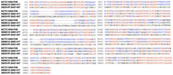
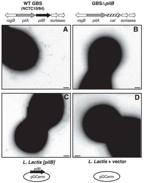
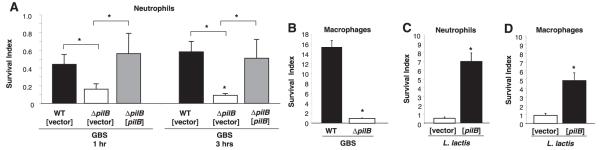
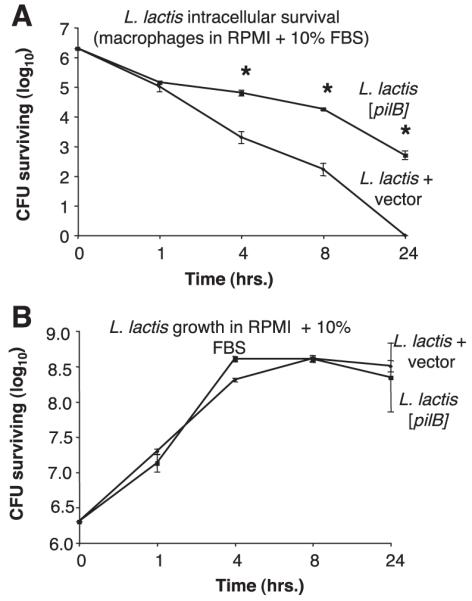
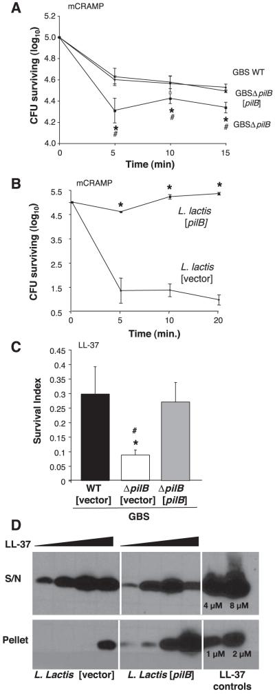
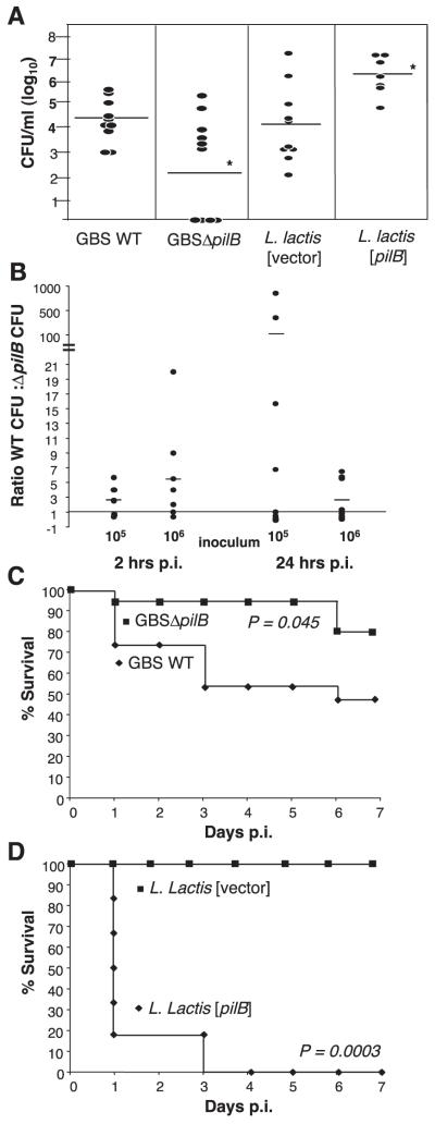
Group B Streptococcus (GBS) is a major cause of invasive bacterial infections in newborns and certain adult populations. Surface filamentous appendages known as pili have been recently identified in GBS. However, little is known about the role of these structures in disease pathogenesis. In this study we sought to probe potential functional role(s) of PilB, the major GBS pilus protein subunit, by coupling analysis of an isogenic GBS pilB knockout strain with heterologous expression of the pilB gene in the nonpathogenic bacterium Lactococcus lactis. We found the knockout GBS strain that lacked PilB was more susceptible than wild-type (WT) GBS to killing by isolated macrophages and neutrophils. Survival was linked to the ability of PilB to mediate GBS resistance to cathelicidin antimicrobial peptides. Furthermore, the PilB-deficient GBS mutant was more readily cleared from the mouse bloodstream and less-virulent in vivo compared to the WT parent strain. Strikingly, overexpression of the pilB gene alone in L. lactis enhanced resistance to phagocyte killing, increased bloodstream survival, and conferred virulence in a mouse challenge model. Together these data demonstrate that the pilus backbone subunit, PilB, plays an integral role in GBS virulence and suggests a novel role for gram-positive pili in thwarting the innate defenses of phagocyte killing.


Figures

Figure 1
Figure 1
GBS PilB protein sequence alignment. Amino acid sequences and alignment of NCTC10/84 PilB and the homologous pili subunit proteins SAG1407 and GBS14778 expressed by the two sequenced GBS strains, NEM316 and 2603 V/R, respectively. Residues colored in red are identical in all three strains, while blue residues indicate identity in only two of the three GBS strains and green residues share similarity but are not identical.
Figure 2
Figure 2
GBS PilB surface expression on GBS and L. lactis. Immunogold labeling and transmission electron microscopy (TEM) with mouse sera against GBS59 (PilB) for WT GBS (A), GBSΔpilB (B), L. lactis expressing pilB (C), and WT L. lactis containing vector only (D). Scale bars = 100 nm.
Figure 3
Figure 3
GBS PilB promotes resistance to neutrophil and macrophage clearance. Survival index of GBS WT [vector], GBSΔpilB [vector], and GBSΔpilB [pilB] on coculture with human neutrophils at 1 and 3 h, MOI = 10 (A). Survival index of GBS WT and GBSΔpilB in coculture with murine peritoneal macrophages for 3 h, MOI = 5(B). Survival index of WT L. lactis with vector only and expressing pilB in coculture with human neutrophils for 2 h at MOI = 5 (C) and coculture with murine peritoneal macrophages for 2 h, MOI = 1 (D). All data shown are a representative experiment of at least 3 experiments. Error bars represent the 95% confidence interval of the mean of 3 wells. Differences in recovered bacteria were analyzed by Student’s t test and are compared to the parent or complemented strain as indicated by brackets (*P<0.05).
Figure 4
Figure 4
Expression of GBS PilB promotes intracellular L. lactis survival in macrophages. Intracellular survival of WT L. lactis containing vector alone or [pilB] during coculture with murine peritoneal macrophages at MOI = 5 in RPMI 1640 cell culture media supplemented with 10% FBS (A). Growth of WT L. lactis containing vector alone or [pilB] in RPMI 1640 cell culture media supplemented with 10% FBS (B). All data shown are a representative experiment of at least 3 experiments. Error bars represent the 95% confidence interval of the mean of 3 wells. Differences in recovered bacteria were analyzed by Student’s t test and are compared to the parent stain (*P<0.05).
Figure 5
Figure 5
GBS PilB confers resistance to antimicrobial peptides. Killing kinetics of WT GBS, GBSΔpilB, and GBSΔpilB [pilB] (A) and WT L. lactis containing vector and L. lactis [pilB] on exposure to 16 μM mCRAMP (B). Survival of GBS WT [vector], GBSΔpilB [vector], and GBSΔpilB [pilB] on exposure to 16 μM LL-37 for 2 h (C). Western blot analysis of human cathelicidin (LL-37) association with bacterial cell surface using WT L. lactis with vector only and L. lactis [pilB] strains (D). All data shown are a representative experiment of at least 3 experiments. Error bars represent the 95% confidence interval of the mean of 3 wells. Differences in recovered bacteria were analyzed by Student’s t test and are compared to the parent stain (*P<0.05) or the complemented strain (#P<0.05).
Figure 6
Figure 6
GBS PilB expression is necessary and sufficient for bacterial virulence in vivo. CFUs from blood collected 24 h post-i.v. injection with 107 CFU/mouse of WT GBS and GBSΔpilB or 4 × 10⁸ CFU/mouse of WT L. lactis containing vector only and L. lactis [pilB] (A). Ratio of GBS WT CFUs to GBSΔpilB CFUs recovered from blood collected 2 and 24 h following i.v. coinjection with 105 or 106 CFU/mouse of each strain. Each circle represents one mouse (B). Kaplan-Meyer survival curve of mice injected i.p., 5 × 10⁷ CFU/mouse, WT GBS or GBSΔpilB (n=15) (C). Survival curve of mice injected i.p., 3.2 × 10⁷ CFU/mouse, L. lactis containing vector only or L. lactis [pilB] (n=6) (D).

Similar articles

Cited by

References

    1. Doran KS, Nizet V. Molecular pathogenesis of neonatal group B streptococcal infection: no longer in its infancy. Mol. Microbiol. 2004;54:23–31. - PubMed
    1. Lauer P, Rinaudo CD, Soriani M, Margarit I, Maione D, Rosini R, Taddei AR, Mora M, Rappuoli R, Grandi G, Telford JL. Genome analysis reveals pili in group b streptococcus. Science. 2005;309:105. - PubMed
    1. Mora M, Bensi G, Capo S, Falugi F, Zingaretti C, Manetti AG, Maggi T, Taddei AR, Grandi G, Telford JL. Group A Streptococcus produce pilus-like structures containing protective antigens and lancefield t antigens. Proc. Natl. Acad. Sci. U. S. A. 2005;102:15641–156466. - PMC - PubMed
    1. Barocchi MA, Ries J, Zogaj X, Hemsley C, Albiger B, Kanth A, Dahlberg S, Fernebro J, Moschioni M, Masignani V, Hultenby K, Taddei AR, Beiter K, Wartha F, von Euler A, Covacci A, Holden DW, Normark S, Rappuoli R, Henriques-Normark B. A pneumococcal pilus influences virulence and host inflammatory responses. Proc. Natl. Acad. Sci. U. S. A. 2006;103:2857–2862. - PMC - PubMed
    1. Rosini R, Rinaudo CD, Soriani M, Lauer P, Mora M, Maione D, Taddei A, Santi I, Ghezzo C, Brettoni C, Buccato S, Margarit I, Grandi G, Telford JL. Identification of novel genomic islands coding for antigenic pilus-like structures in Streptococcus agalactiae. Mol. Microbiol. 2006;61:126–141. - PubMed

Publication types

LinkOut - more resources