. 2008 Jan 15;22(2):212-25.
doi: 10.1101/gad.1601808.

The dynamic interplay between a cell fate determinant and a lysozyme homolog drives the asymmetric division cycle of Caulobacter crescentus

Affiliations

The dynamic interplay between a cell fate determinant and a lysozyme homolog drives the asymmetric division cycle of Caulobacter crescentus

Sunish Kumar Radhakrishnan et al. Genes Dev. .

Abstract

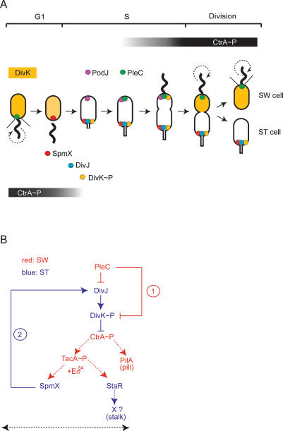
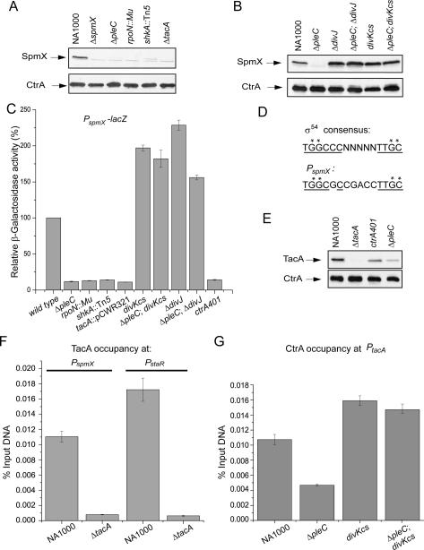
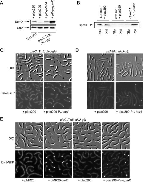
Caulobacter crescentus divides asymmetrically into a swarmer cell and a stalked cell, a process that is governed by the imbalance in phosphorylated levels of the DivK cell fate determinant in the two cellular compartments. The asymmetric polar localization of the DivJ kinase results in its specific inheritance in the stalked daughter cell where it phosphorylates DivK. The mechanism for the polar positioning of DivJ is poorly understood. SpmX, an uncharacterized lysozyme homolog, is shown here to control DivJ localization and activation. In the absence of SpmX, DivJ is delocalized and dysfunctional, resulting in developmental defects caused by an insufficiency in phospho-DivK. While SpmX stimulates DivK phosphorylation in the stalked cell, unphosphorylated DivK in the swarmer cell activates an intricate transcriptional cascade that leads to the production of the spmX message. This event primes the swarmer cell for the impending transition into a stalked cell, a transition that is sparked by the abrupt accumulation and localization of SpmX to the future stalked cell pole. Localized SpmX then recruits and stimulates DivJ, and the resulting phospho-DivK implements the stalked cell fate. The dynamic interplay between SpmX and DivK is at the heart of the molecular circuitry that sustains the Caulobacter developmental cycle.


Figures

Figure 1.
Figure 1.
Graphical representation of the cell fate regulators in Caulobacter and the genetic circuit they constitute. (A) Subcellular localization of DivJ, DivK, PleC, and the localization factors PodJ and SpmX during the Caulobacter asymmetric division cycle: SpmX (red dot) localizes to the pole previously occupied by PleC (green dot), which eventually morphs into the stalked pole. SpmX recruits DivJ (blue dot), initiating the phosphorylation and localization of DivK (yellow), eventually resulting in bipolar DivK∼P (yellow dot) in predivisional cells. Predivisional cells also display PleC at the flagellated pole. PleC has been recruited previously to that site by PodJ (purple dot), which localizes to that pole in late stalked cells (Viollier et al. 2002b; Hinz et al. 2003). Coincident with the compartmentalization of the late predivisional cell, localized DivK∼P and diffuse (dephosphorylated) DivK are present in the stalked (ST) cell chamber and the swarmer (SW) cell chamber, respectively. Pili and the flagellum are indicated by the thin lines (black) and the thick wavy line, respectively. The circular dashed arrow denotes a rotating flagellum. The graded black bar indicates the time during the cell cycle that CtrA∼P is present. In late predivisional cells, CtrA∼P accumulates in the swarmer compartment and is eliminated from the stalked cell compartment. (B) In the swarmer cell compartment (SW, red), PleC reduces DivK∼P levels by direct dephosphorylation of DivK∼P (rectangular arrow, event 1) (Matroule et al. 2004), and possibly indirectly by inhibiting the DivJ kinase (Sommer and Newton 1991; Wheeler and Shapiro 1999) by the production of an inhibitor or modulator of DivJ activity. High levels of DivK∼P inhibit CtrA∼P-mediated transcriptional activation of tacA and pilA by a mechanism that is poorly understood (Biondi et al. 2006a). The tacA translation product along with σ54-containing RNA polymerase (Eσ54) catalyzes transcription of spmX and staR, the gene for a transcriptional regulator of stalk biogenesis (Biondi et al. 2006b). At the swarmer-to-stalked cell transition, SpmX accumulates to localize and activate DivJ (event 2), thereby producing a surge in DivK∼P that signals the removal of CtrA∼P by a complex phosphosignaling cascade (Biondi et al. 2006a; Iniesta et al. 2006). This event, coupled with the disappearance of TacA at the swarmer-to-stalked cell transition (see Fig. 4A), leads to the shut-down of SpmX transcription in stalked cells (ST, blue). The dotted lines indicate transcriptional regulation; bold lines indicate the regulation at the level of protein localization or activity. The black dashed arrow indicates the new connections of the circuit uncovered herein.
Figure 2.
Figure 2.
A motility mutant screen identifies spmX, an uncharacterized gene encoding a muramidase homolog required for proper cell division. (A) Domain organization of SpmX showing the predicted transmembrane domains (brown), the muramidase domain (yellow), the position of the Himar1 (white triangle), and the EZ-Tn5 (black triangle) insertion in strains NS349 and NS190, respectively. The line below the domain architecture shows the deleted coding region of the ΔspmX strain. (B) Motility assay of NA1000 (wild type), NS190, NS349, ΔspmX, ΔpleC, and ΔdivJ strains. Overnight cultures (2.5 μL) were placed on PYE swarm (0.3%) agar plates and incubated for 60 h at 30°C. Motility defects can be seen as swarms with a compact appearance, whereas those from wild type are diffuse and enlarged. (C–G) Transmission electron micrographs of wild-type (E), ΔspmX (D,F,G), and divKcs (C) cells grown at 30°C. Pili (visualized indirectly by staining with pilus-specific bacteriophage ΦCbK) and flagella are indicated by white and black arrows, respectively. Arrowheads indicate bipolar stalks (black) or abnormal constrictions (white). Bars, ∼200 nm.
Figure 3.
Figure 3.
Localization and in vivo phosphorylation of DivK and DivJ are impaired in the ΔspmX mutant. (A,B) Differential interference contrast (DIC) and fluorescence micrographs of NA1000, ΔspmX, and ΔspmX ΔpleC cells expressing either DivK-GFP expressed from a low-copy plasmid (pdivK-gfp) under the control of the native divK promoter (A) or DivJ-YFP from the endogenous divJ locus (B). (C) Determination of relative DivK∼P/DivK levels in NA1000, ΔspmX, ΔpleC ΔspmX, ΔpleC, and ΔdivJ cells. (D) Measurement of relative DivJ∼P/DivJ levels in NA1000 and ΔspmX cells. Bars, ∼2 μm.
Figure 4.
Figure 4.
Colocalization of SpmX and DivJ. (A) Immunoblots showing cellular levels of SpmX, DivJ, PilA, TacA, and CtrA during various stages of the wild-type (NA1000) cell cycle. The orange bars indicate the time when spmX mRNA is present during the cell cycle as determined previously (Laub et al. 2000; McGrath et al. 2007). (B) Polar localization of SpmX-mCherry and mCherry-SpmX, expressed from the endogenous chromosomal locus in place of wild-type SpmX in strains MT237 and MT272, respectively. (C) Immunofluorescence micrographs using anti-SpmX antiserum in LS3200 (divJ-gfp) cells. SpmX-derived signals (red) colocalize (yellow) with DivJ-GFP signals (green). Cells were visualized by staining in 1 μg/mL DAPI (4′,6-diamidino-2-phenylindole). No SpmX-derived foci were seen at the poles in ΔspmX cells. (D) DivJ-YFP (green) and mCherry-SpmX (red) colocalize (yellow) to the stalked pole of NR3601 (spmX-mCherry; divJ-yfp) cells in which the modified alleles replaced the native spmX and divJ genes. (E) Time-lapse fluorescence microscopy of purified NR3601 swarmer cells growing on a cushion of 1% M2G agarose on a microscope slide, showing that SpmX-mCherry (red arrowheads) localizes to the pole first, followed by that of DivJ-YFP (yellow arrowheads), and finally the stalk (white arrow heads) elongates from the same pole. Bars, ∼2 μm. (F) Immunoprecipitation (IP) analysis of membrane-solubilized extracts of the divJ-yfp and NA1000 strain with a monoclonal antibody to GFP (α-GFP). Precipitated samples were analyzed by immunoblotting (Blot) using specific polyclonal antibodies to SpmX (α-SpmX). (G) Immunoblot analysis of extracts from NA1000 and divJ-yfp to detect DivJ-YFP using a monoclonal antibody to GFP. The asterisk (*) marks two nonspecific signals reacting with the anti-GFP antibody. (H) Immunoprecipitation experiments using membrane solubilized extracts of the divJ-yfp and ΔspmX; divJ-yfp strains and polyclonal antibodies to SpmX. DivJ-YFP was detected by immunoblotting using a monoclonal antibody to GFP. Note that the two nonspecific signals (*) do not coprecipitate. The white arrowhead marks the position of the signals derived from the IgG heavy chains.
Figure 5.
Figure 5.
Role of the muramidase domain in the localizing of SpmX and DivJ to the stalked pole. (A) Fluorescence and DIC micrographs of ΔspmX; divJ-yfp cells expressing SpmX derivatives from a vanillate-inducible promoter (Pvan) on low-copy plasmid pRVMCS-5 (Thanbichler et al. 2007) after growth in PYE supplemented with tetracycline (1 μg/mL) and induced with vanillate (50 nM) for 2 h. (B) Immunoblot analysis of SpmX and CtrA steady-state levels present in equal amounts of cellular extracts from strains in A. Note that the SpmX(1–150) derivative is not efficiently detected by the anti-SpmX antibody because a recombinant SpmX derivative from residues 120–351 was used as immunogen. (C) Localization of SpmX-mCherry derivatives expressed from pRVMCS-5 in NA1000 cells grown as in A. Note that as a result of ectopic expression from Pvan, SpmX-mCherry can often be seen to localize to the pole opposite the stalk. (D) Immunoblot experiments to determine SpmX-mCherry steady-state levels present in cell extracts from strains in C. Equal amounts of cell extracts were loaded in each lane. Similar localization results and abundance patterns as in C and D, respectively, were observed when the SpmX-mCherry derivatives were expressed in a ΔspmX background (data not shown). (E) NA1000 cells expressing the SpmX-mCherry or SpmX(1–350)-mCherry (as in D) were converted to spheroblasts (SB) and gently centrifuged. The cell (SB Cells) and supernatant (SB Sup.) fraction, along with a control lysate of untreated cells (UT Lysate), were examined for the presence of SpmX-mCherry or CtrA by immunoblotting. The presence of SpmX(1– 350)-mCherry in the spheroblast supernatant indicates that it is exported into the periplasm in untreated cells. Note that spheroblasting is incomplete, explaining why a substantial amount of SpmX(1–350)-mCherry remains associated with cells. Similar experiments with SpmX(1–150)-mCherry were inconclusive because spheroblasts were fragile, undergoing lysis during centrifugation (data not shown). (F) The immunoblot shown in E was reprobed for the presence of a cytoplasmic protein (CtrA) as a control for cell lysis. In B, D, E, or F, SpmX-mCherry derivatives and CtrA were detected using polyclonal antibodies to SpmX (α-SpmX), CtrA (α-CtrA), and mRFP (α-mRFP). Note that the anti-mRFP antibody reacts with mCherry.
Figure 6.
Figure 6.
Transcriptional regulation of spmX. (A) Immunoblots showing SpmX steady-state levels in wild-type (NA1000), ΔpleC, rpoN∷Tn5, shkA∷Tn5, and ΔtacA mutants. (B) Immunoblots showing that ΔdivJ and divKcs mutations restore SpmX production to ΔpleC mutant cells. (C) β-Galactosidase assays using the PspmX-lacZ reporter plasmid to determine spmX promoter activity in NA1000 and various mutants. Temperature-sensitive mutants were grown at the permissive temperature. (D) Comparison of the spmX promoter sequence with the Eσ54 consensus sequence. Asterisks (*) indicate nucleotides required for Eσ54 binding. Underlined are the −24 and −12 consensus sequences for Eσ54 promoters. (E) Immunoblot showing TacA steady-state levels in NA1000, ΔtacA, ctrA401, and ΔpleC mutants. CtrA was used as a loading control for the immunoblots in A, B, and E. (F) Measurement of TacA occupancy at the spmX and staR promoters in vivo using qChIP analysis. (G) qChIP experiments showing the reduction in CtrA occupancy at the tacA promoter in ΔpleC mutant cells, and an increase in the divKcs single mutant and the ΔpleC divKcs double mutant relative to NA1000.
Figure 7.
Figure 7.
The absence of SpmX underlies the DivJ localization defect in pleC and ctrA401 mutants. (A,B) Immunoblots of pleC∷Tn5 divJ-gfp and ctrA401 divJ-gfp cells upon expression of TacA or SpmX from a xylose-inducible promoter on a low-copy plasmid (pPxyl-tacA). In A, cells were grown in PYE containing tetracycline (1 μg/mL) and 20 mM xylose, except for the strain containing pPxyl-spmX that was grown in 2 mM xylose and tetracycline. In B, cells were grown in PYE containing tetracycline (1 μg/mL) as well as 20 mM xylose (Xyl) or 20 mM glucose (Glu). (C,D) Fluorescence micrographs showing DivJ-GFP in pleC∷Tn5 divJ-gfp (C) and ctrA401 divJ-gfp (D) cells that were grown in PYE containing tetracycline (1 μg/mL) and 20 mM xylose and harbored either plasmid pPxyl-tacA or the empty vector. (E) Localization of DivJ-GFP in a pleC∷Tn5 mutant that expresses SpmX from a xylose-inducible promoter on a low-copy plasmid (pPxyl-spmX) or with a complementing plasmid harboring the pleC gene. No localization is observed in strains with the empty vector. Cells were grown in PYE containing tetracycline (1 μg/mL). Bars, ∼2 μm.

References

    1. Alley M.R., Maddock J.R., Shapiro L. Polar localization of a bacterial chemoreceptor. Genes & Dev. 1992;6:825–836. - PubMed
    1. Biondi E.G., Reisinger S.J., Skerker J.M., Arif M., Perchuk B.S., Ryan K.R., Laub M.T. Regulation of the bacterial cell cycle by an integrated genetic circuit. Nature. 2006a;444:899–904. - PubMed
    1. Biondi E.G., Skerker J.M., Arif M., Prasol M.S., Perchuk B.S., Laub M.T. A phosphorelay system controls stalk biogenesis during cell cycle progression in Caulobacter crescentus. Mol. Microbiol. 2006b;59:386–401. - PubMed
    1. Chen J.C., Viollier P.H., Shapiro L. A membrane metalloprotease participates in the sequential degradation of a Caulobacter polarity determinant. Mol. Microbiol. 2005;55:1085–1103. - PubMed
    1. Chen J.C., Hottes A.K., McAdams H.H., McGrath P.T., Viollier P.H., Shapiro L. Cytokinesis signals truncation of the PodJ polarity factor by a cell cycle-regulated protease. EMBO J. 2006;25:377–386. - PMC - PubMed

Publication types

LinkOut - more resources