. 2008 Jan;6(1):e6.
doi: 10.1371/journal.pbio.0060006.

Conformational equilibria in monomeric alpha-synuclein at the single-molecule level

Affiliations

Conformational equilibria in monomeric alpha-synuclein at the single-molecule level

Massimo Sandal et al. PLoS Biol. 2008 Jan.

Abstract

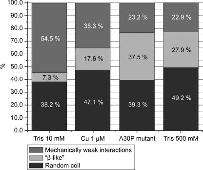
Human alpha-Synuclein (alphaSyn) is a natively unfolded protein whose aggregation into amyloid fibrils is involved in the pathology of Parkinson disease. A full comprehension of the structure and dynamics of early intermediates leading to the aggregated states is an unsolved problem of essential importance to researchers attempting to decipher the molecular mechanisms of alphaSyn aggregation and formation of fibrils. Traditional bulk techniques used so far to solve this problem point to a direct correlation between alphaSyn's unique conformational properties and its propensity to aggregate, but these techniques can only provide ensemble-averaged information for monomers and oligomers alike. They therefore cannot characterize the full complexity of the conformational equilibria that trigger the aggregation process. We applied atomic force microscopy-based single-molecule mechanical unfolding methodology to study the conformational equilibrium of human wild-type and mutant alphaSyn. The conformational heterogeneity of monomeric alphaSyn was characterized at the single-molecule level. Three main classes of conformations, including disordered and "beta-like" structures, were directly observed and quantified without any interference from oligomeric soluble forms. The relative abundance of the "beta-like" structures significantly increased in different conditions promoting the aggregation of alphaSyn: the presence of Cu2+, the pathogenic A30P mutation, and high ionic strength. This methodology can explore the full conformational space of a protein at the single-molecule level, detecting even poorly populated conformers and measuring their distribution in a variety of biologically important conditions. To the best of our knowledge, we present for the first time evidence of a conformational equilibrium that controls the population of a specific class of monomeric alphaSyn conformers, positively correlated with conditions known to promote the formation of aggregates. A new tool is thus made available to test directly the influence of mutations and pharmacological strategies on the conformational equilibrium of monomeric alphaSyn.


Conflict of interest statement

Competing interests. The authors have declared that no competing interests exist.

Figures

Figure 1
Figure 1. The Mechanical Signatures of αSyn Conformational Classes as Recorded by SMFS
(A) Schematic representation of the polyprotein constructs used in this work: 3S3 contains the αSyn sequence (red) flanked on either side by three titin I27 modules (blue), the N-terminal His-tag needed for purification purposes (green), and the C-terminal Cys-Cys tail needed for covalent attachment to the gold surface (yellow). In 1S1, the αSyn moiety is flanked only by one I27 on both sides; the 3T is made up by three I27s. In the αSyn moiety (enlarged), three regions are shown: (i) the amphipathic region, prone to fold in α-helical structures when in contact with phospholipid membranes; (ii) the fibrillogenic NAC region, characteristic of the fibril core of αSyn amyloid; and (iii) the acidic C-terminal tail, strongly charged and not prone to fold. The positions of alanine 30, site of the A30P mutation and histidine 50, which is crucial for the binding of Cu2+, are marked. (B) Example of curve characterized by a featureless region assigned to the stretching of αSyn moiety having, in this case, the mechanical properties of a random coil (see Results section). This region is followed (from left to right) by six unfolding peaks of about 200 pN, with about 28-nm gaps between each, assigned to the unfolding of I27 domains. (C) Example of the curves featuring the β-like signature of αSyn (see Results section), showing seven practically indistinguishable unfolding events of similar magnitude and spacing. (D) Curves featuring the signature of mechanically weak interactions, showing single or multiple small peaks (arrows) superimposed on the purely entropic WLC behavior of the trace preceding the six saw-tooth–like peaks. The right panels show a zoom of the region enclosed by the dashed squares.
Figure 2
Figure 2. Population Shift of αSyn Conformers in Different Conditions
Population of αSyn conformers in the four different conditions tested in the present work. Percentages observed for each curve type (see Figure 1) at 10 mM Tris/HCl (n = 55), 10 mM Tris/HCl with 1 μM Cu2+ (n = 34), the A30P mutant in 10 mM Tris/HCl (n = 56), and 500 mM Tris/HCl (n = 61).
Figure 3
Figure 3. Contour Length Analysis of β-Like Force Curves
Values of the first peak position in force curves showing seven unfolding peaks. The height of each bar corresponds to the initial contour length of a single curve, obtained by fitting the first unfolding peak by means of the WLC model. The dashed line is the length corresponding to a protein construct with six I27 folded modules plus 95 aa of αSyn folded into a β-like structure, and the remaining 50 aa of αSyn unstructured (see Discussion section). The dashed-and-dotted line is the length corresponding to a protein construct with six I27 modules plus the 140 aa of αSyn completely unstructured. The lengths of twelve randomly chosen I27 modules have also been reported (dark gray columns) for comparison. The solid line is the nominal I27 contour length. The larger spreading of the αSyn data confirms the higher conformational heterogeneity. Side quotas show the difference between the maximum and minum observed length value for I27 (bottom) and β-like structures (top).
Figure 4
Figure 4. Circular Dichroism and Fluorescence Spectroscopy
(A) Fluorescence spectra of αSyn, 3T, and 1S1 (dotted, dashed, and solid line, respectively). (B) Circular dichroism spectra in PBS buffer of 3T, 1S1 (solid lines). The αSyn contribution in 1S1 (dashed line) is calculated by subtracting the relative contribution of the I27 domains from the CD spectrum of 1S1. (C) CD spectra of αSyn in PBS (solid line) and 250 mM SDS (dotted line). The αSyn contribution in 1S1 (dashed line) is reported as in (B).

References

    1. Fink AL. Natively unfolded proteins. Curr Opin Struct Biol. 2005;15:35–41. - PubMed
    1. Dyson HJ, Wright PE. Intrinsically unstructured proteins and their functions. Nat Rev Mol Cell Biol. 2005;6:197–208. - PubMed
    1. Chiti F, Dobson CM. Protein misfolding, functional amyloid, and human disease. Annu Rev Biochem. 2006;75:333–366. - PubMed
    1. Cookson MR. Hero versus antihero: the multiple roles of alpha-synuclein in neurodegeneration. Exp Neurol. 2006;199:238–242. - PubMed
    1. Uversky VN, Fink AL. Protein misfolding, aggregation and conformational diseases. New York: Kluwer Academic/Plenum Publishers; 2006.

Publication types

Associated data