

doi: 10.1016/j.immuni.2007.11.023.
The lupus-related Lmb3 locus contains a disease-suppressing Coronin-1A gene mutation
Affiliations
- PMID: 18199416
- PMCID: PMC2274909
- DOI: 10.1016/j.immuni.2007.11.023
Item in Clipboard
The lupus-related Lmb3 locus contains a disease-suppressing Coronin-1A gene mutation
Immunity.
2008 Jan.
Abstract
Here, we show that a lupus-suppressing locus is caused by a nonsense mutation of the filamentous actin-inhibiting Coronin-1A gene. This mutation was associated with developmental and functional alterations in T cells including reduced migration, survival, activation, and Ca2+ flux. T-dependent humoral responses were impaired, but no intrinsic B cell defects were detected. By transfer of T cells, it was shown that suppression of autoimmunity could be accounted for by the presence of the Coro1a(Lmb3) mutation in T cells. Our results demonstrate that Coronin-1A is required for the development of systemic lupus and identify actin-cytoskeleton regulatory proteins as potential targets for modulating autoimmune diseases.
Figures
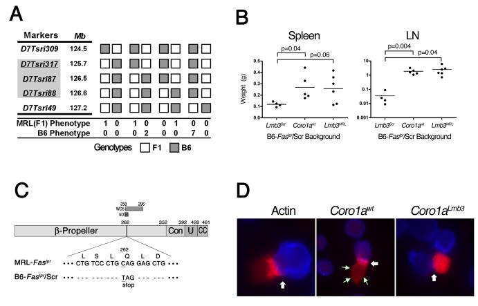
(A) Fine mapping of Lmb3. The interval (indicated by shaded markers) was reduced to 0.9 Mb between D7Tsri317 and D7Tsri88 using ∼2500 meioses (see Methods). Only crossovers between D7Tsri309 and T7Tsri49 are shown. MRL and B6 phenotypes were determined by numbers of CD4+ and DN LN T cells 5 wk after subcutaneous CFA injection. (B) B6-Faslpr/Scr.B6-Coro1awt mice express the Lmb3 phenotype. Wet weights of spleen and LN from 11-12 month-old wild-type or congenic B6-Faslpr/Scr mice that have either the original Lmb3 interval (Lmb3Scr), the Coro1awt from the B6/J, or the MRL Lmb3 interval (Lmb3MRL). (C) Coro1aLmb3 Q262X allele. A nonsense C→T transition at residue 784 changes a glutamine (CAG) to a termination codon (TAG) within the 5th WD40 domain (5D region of the β-propeller domain). Resulting polypeptide lacks a significant portion of the β-propeller, as well as the entire distal constant (Con), unique (U) and coiled coil (CC) regions. (D) Coro1aLmb3 protein fails to localize to the plasma membrane and immune synapse. Jurkat cells were transfected with plasmids encoding C-terminal-fluorescent protein-labeled actin, Coro1awt or Coro1aLmb3 and mixed with SED-pulsed Cy-5-labeled Raji antigen presenting cells (100X). Immune synapse (white arrows) and plasma membrane Coro1a (green arrows) are indicated. Data representative of two transfection experiments.
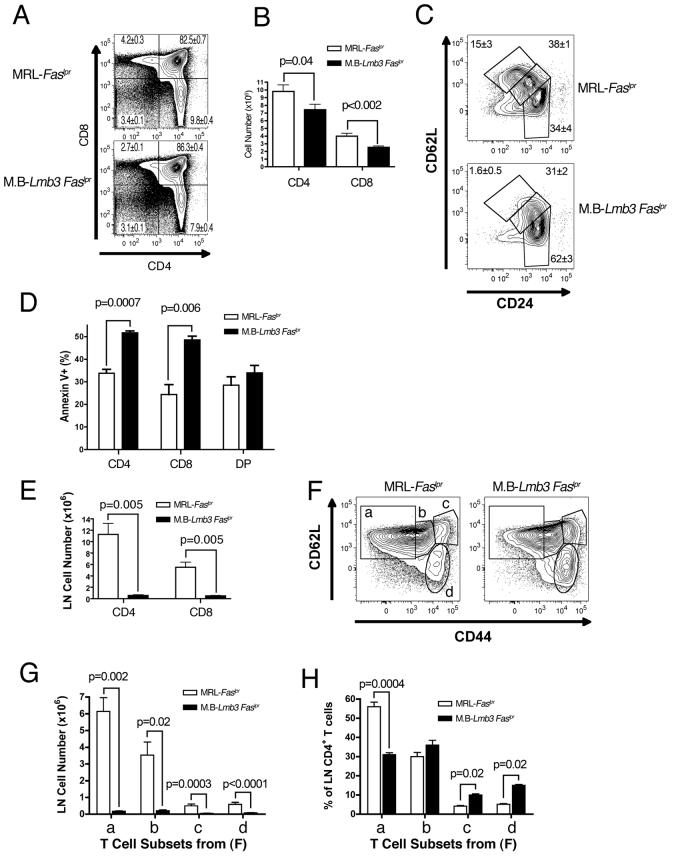
(A) Reduction in percentages of thymic CD4+ and CD8+ single positive (SP) T cells. Mean±SEM of 4-7 wk.-old mice, n=15 (MRL-Faslpr), 11 (M.B-Lmb3 Faslpr). A representative profile is shown. (B) Thymic SP T cell numbers are reduced in MRL.B6-Lmb3 Faslpr mice. n=11-12/group. (C) Reduction in mature (CD24lo, 62Lhi) subset of CD4+ SP thymocytes. Representative data from 4-5 mice/group (see Figure S1 for CD8+ SP subset and single markers). (D) Increased spontaneous cell death in SP thymocytes. n=3-4/group. (E) Reduced numbers of CD4+ and CD8+ LN T cells. n=8-15/group. (F) LN CD44 and CD62L-defined CD4+ T cell subsets. (G) Reduced numbers of all CD4+ T cell subsets defined in panel F. (H) Reduced percentages of the CD44lo CD62Lhi (naïve) subset defined in panel F. Data for (FH) from 3-4/group. (see Figure S2 for CD4+ T cell subsets in the spleen and CD8+ T cell subsets in the LN and spleen).
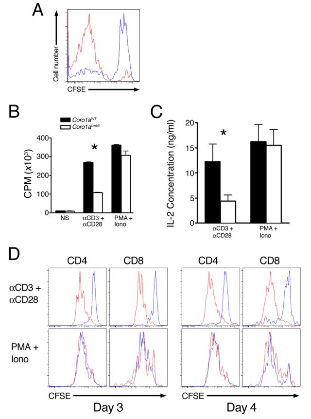
(A) Reduction in lymphopenia-induced homeostatic proliferation. CFSE-labeled B6 (red line) or B6-Coro1aLmb3 (blue line) Thy1.2 T cells were transferred into sublethally irradiated Thy1.1-recipient B6.PL mice and CFSE-staining of Thy1.1-negative LN CD4+ T cells was analyzed by flow cytometry 5 d later. n=3 mice/group. (B) Reduced MRL.B6-Lmb3 Faslpr T cell proliferation to anti-CD3/anti-CD28 (P<0.05), but not PMA/Ionomycin. (C) IL-2 production is lower in MRL-Faslpr T cells with Coro1aLmb3 (p<0.05). Purified T cells from MRL-Faslpr or MRL.B6-Lmb3 Faslpr mice were activated with anti-CD3/CD28 or PMA/Ionomycin for 48 hrs and supernatants analyzed for IL-2 production. (D) Reduced division of Coro1aLmb3 mutant T cells after stimulation with anti-CD3/anti-CD28, but not PMA/Ionomycin. Coro1awt (red lines), Coro1aLmb3 (blue lines). Data are representative of 3 independent experiments.
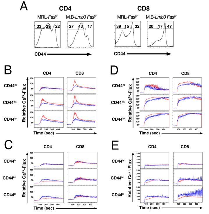
(A) CD44 low, intermediate and high subsets of CD4+ and CD8+ T from Coro1awt and Coro1aLmb3 mice. (B) Ca2+-flux in CD44 low, intermediate and high subsets of CD4+ and CD8+ T cells in the presence of exogenous calcium following TCR engagement (anti-CD3/anti-CD38 antibodies). (C) Ca2+-flux in CD44 low, intermediate and high subsets of CD4+ and CD8+ T cells in the absence of exogenous calcium following TCR engagement. (D) Ca2+-flux in CD44 low, intermediate and high subsets of CD4+ and CD8+ T cells in the presence of exogenous calcium following Ionomycin stimulation. (E) Ca2+-flux in CD44 low, intermediate and high subsets of CD4+ and CD8+ T cells in the absence of exogenous calcium following Ionomycin stimulation. Coro1awt (red lines), Coro1aLmb3 (blue lines). Data are representative of 3 independent experiments.
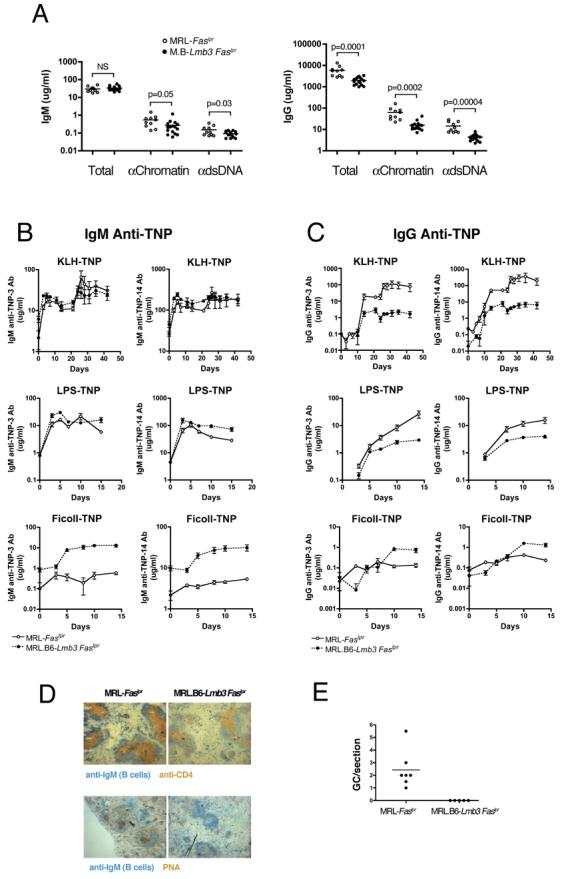
(A) IgG polyclonal (total) and anti-nuclear antibodies from 4 mo. old mice (right panel) are reduced to a greater degree than IgM antibodies (left panel). (B-C) IgM and IgG TNP levels to TNP-14 (low and high affinity antibodies) and TNP-3 (high affinity antibodies) after immunization with T-D (TNP-KLH), T-I type 1, (TNP-LPS), or T-I type 2 (TNP-Ficoll) antigens. n=5-6 (T-D) or 9-12 (T-I). T-I groups were immunized on day 0 and T-D groups on day 0 and 21. (D) Representative spleen sections stained for CD4+ T cells, B cells (IgM+), and GC (PNA+) 10 days after i.p. TNP-KLH in CFA. (E) Number of GC per spleen section 10 d after i.p. TNP-KLH in CFA. n= 5 for congenic and 7 for wild-type MRL-Faslpr mice (D,E).
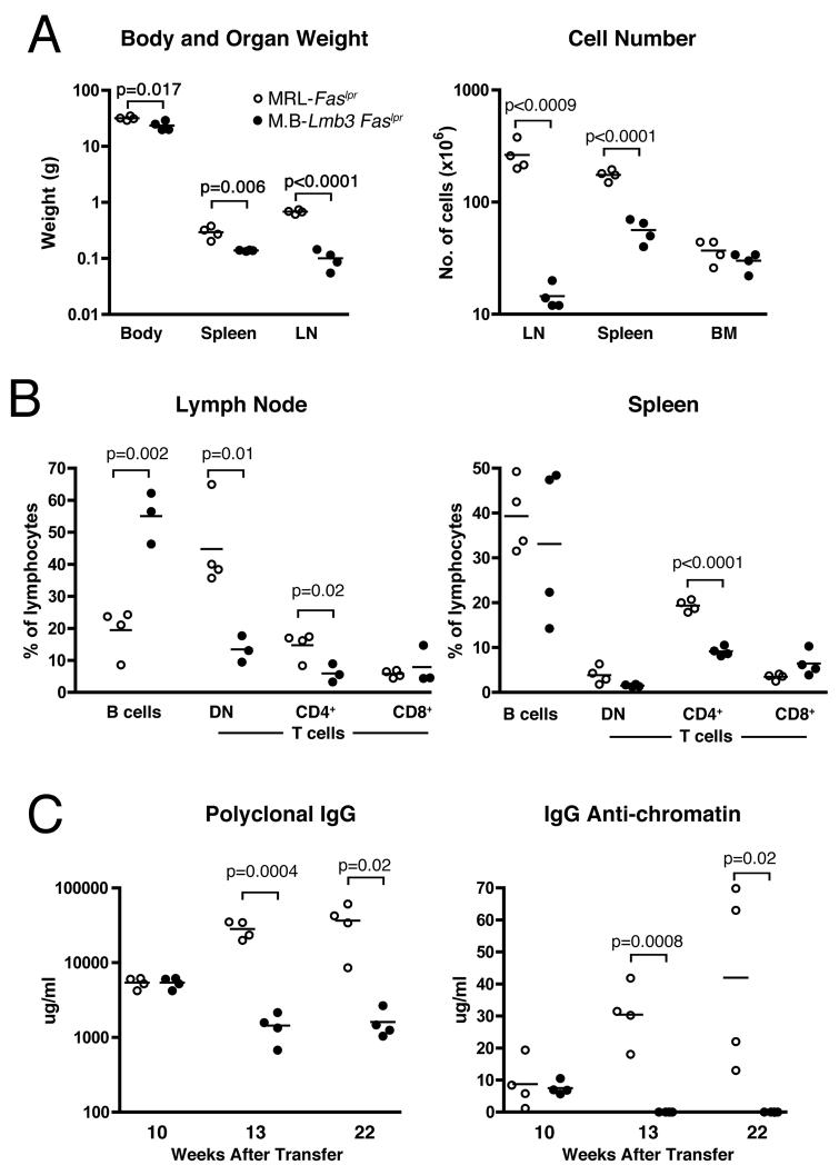
Purified LN T cells from MRL-Faslpr or MRL.B6-Lmb3 Faslpr (M.B-Lmb3 Faslpr) were transferred into T cell receptor β-chain-deficient MRL-Faslpr mice. Lupus-related traits in panels A and B were determined 22 wk. after transfer. (A) Body and lymphoid organ weights. Total cell numbers of lymphoid organs. (B) Percentages of B cells and T cell subsets in spleen and LN. (C) Total and anti-chromatin IgG serum levels.
Comment in
-
Understanding lupus: fishing genes out of mice and men.Immunity. 2008 Jan;28(1):8-10. doi: 10.1016/j.immuni.2007.12.007. Immunity. 2008. PMID: 18199411
References
-
- Appleton BA, Wu P, Wiesmann C. The crystal structure of murine coronin-1: a regulator of actin cytoskeletal dynamics in lymphocytes. Structure. 2006;14:87–96. - PubMed
-
- Boackle SA, Holers VM, Chen X, Szakonyi G, Karp DR, Wakeland EK, Morel L. Cr2, a candidate gene in the murine Sle1c lupus susceptibility locus, encodes a dysfunctional protein. Immunity. 2001;15:775–785. - PubMed
-
- Dustin ML. Immunology. When F-actin becomes too much of a good thing. Science. 2006;313:767–768. - PubMed
Publication types
MeSH terms
Substances
Grants and funding
LinkOut - more resources
Full Text Sources
Other Literature Sources
Medical
Molecular Biology Databases
Miscellaneous
