. 2008 Jan;28(1):100-11.
doi: 10.1016/j.immuni.2007.11.021.

A two-step process for thymic regulatory T cell development

Affiliations

A two-step process for thymic regulatory T cell development

Chan-Wang Joaquim Lio et al. Immunity. 2008 Jan.

Abstract

Recognition of self-antigens is required for regulatory T (Treg) cells to exert dominant tolerance. However, the mechanism by which self-reactive thymocytes are diverted into the Treg cell subset is unclear. To address this question, we looked for the immediate precursors to Treg cells within Foxp3(-)CD4+CD8(-) thymocytes. By using intrathymic transfer, we found that the CD25hi subset is highly enriched in Treg cell precursors. This was supported by tracking of thymocyte development via analysis of T cell receptor (TCR) repertoires in a TCR-beta transgenic model. These Treg cell precursors exist at a developmental stage where they are poised to express Foxp3 without further TCR engagement, requiring only stimulation by interleukin-2 (IL-2) or IL-15. Thus, we propose that the selection of self-reactive thymocytes into the Treg cell subset occurs via an instructive rather than stochastic-selective model whereby TCR signals result in the expression of proximal IL-2 signaling components facilitating cytokine-mediated induction of Foxp3.


Figures
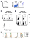
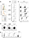
Figure 1
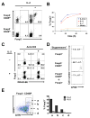
Figure 1. CD25hi Foxp3 CD4SP thymocytes are enriched for Treg cell precursors
(A) Overlap between Foxp3+ and Foxp3 CD4SP thymocytes for the expression of CD25 and GITR. Thymocytes from Foxp3gfp reporter mice were analyzed by flow cytometry. Cells are gated on the CD4SP subset, and are further gated based on Foxp3 (left panel) into the Foxp3 (middle) and Foxp3+ subsets (right). (B) Development of Foxp3+ cells from CD25hi, but not CD25lo, Foxp3 CD4SP cells after intrathymic transfer. FACS purified CD25lo or CD25hi Foxp3 CD45.2+ CD4SP cells (105) were intrathymically injected into congenically marked CD45.1+ recipients. Development of Foxp3+ cells was analyzed by flow cytometry 3 days post transfer. Representative FACS plots for the intrathymically injected CD45.1 CD45.2+ CD4SP cells from two independent experiments are shown on the top, and the percent Foxp3+ cells from individual mice plotted on the bottom. The mean value is indicated by a bar. (C) Maintenance of Foxp3+ cells from CD25hi Foxp3 CD4SP thymic precursors. 105 sorted CD25lo or CD25hi Foxp3 CD4SP thymocytes (Thy1.2+) together with 5x10⁵ MACS purified peripheral CD4+ cells (Thy1.1+) were intravenously transferred into TCRβ−/− mice, and analyzed at 3 and 14 days by flow cytometry of pooled spleen and lymph nodes cells. Representative FACS plots from two independent experiments show cells gated on donor Thy1.2+ Thy1.1 TCRβ+ CD4+ cells. (D) Flow cytometric characterization of CD25hi Foxp3 cells. Thymocytes from Foxp3gfp mice were stained with the indicated antibodies along with CD4, CD8, and CD25; and analyzed on a FACSAria. Histogram plots shown are gated on CD4SP cells.
Figure 2
Figure 2. CD25hi Foxp3 CD4SP TCR repertoire is enriched for Treg cell TCRs
(A) Analysis of shared TCRα sequences among thymic CD4SP populations. Foxp3+, CD25hi GITRhi Foxp3, or CD25lo GITRint Foxp3, CD4SP thymocytes were sorted from TCli TCRβ-transgenic x Foxp3gfp x Tcra+/− mice and TRAV14 (Vα2) TCRα sequences obtained as previously described (Hsieh et al., 2004). Data shown is pooled from two independent experiments (Suppl. Figure 2). Each pie graph represents the TCRα sequences in the data set which were also found in at least one other thymic data set. The portion of the TCRα sequences which overlaps another data set is indicated by a different color in the pie graph, and the data set overlapped is denoted by an arrow. The number of TCRα chains which were not found in another subset was similar between the three subsets (73%, 65%, and 67% for CD25lo, CD25hi, and Foxp3+ subsets, respectively). (B) Comparison of TCRα sequences from thymic and peripheral subsets. Unique TCRα sequences from the indicated thymic CD4SP subsets which were also found in the peripheral TCR data sets are plotted on the x-axis. Each vertical bar represents the likelihood that an individual thymic TCR would be found within the peripheral Foxp3+, naïve (Foxp3 CD44lo), or memory/activated (Foxp3 CD44hi) CD4+ T cell data sets. The size of each color in the bar is calculated by the frequency of the TCR in the subset divided by the sum of the frequencies in all three subsets.
Figure 3
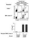
Figure 3. A subset of CD25hi CD4SP thymocytes is at a TCR-independent stage of Treg cell development
CD25lo or CD25hi Foxp3 CD4SP thymocytes (CD45.1+ Thy1.2+) were purified and 105 cells were intravenously transferred along with 5x10⁵ peripheral MACS-purified CD4+ cells (CD45.2+ Thy1.1+) into either TCRβ−/− or MHC class II−/− mice (CD45.2+ Thy1.2+). Development of Foxp3+ T cells from the thymocyte population was assessed 3 days post transfer by flow cytometry. FACS plots are representative of two independent experiments, and are gated on CD45.1+ CD45.2 CD4+ cells. Percentage of cells which are Foxp3+ are plotted below, and shown as mean ± s.d.
Figure 4
Figure 4. Cytokine-mediated induction of Foxp3 in CD25hi Foxp3 CD4SP thymocytes
(A) IL-2 is sufficient to induce Foxp3 in Treg cell precursors in vitro. Sorted CD25lo or CD25hi Foxp3 CD4SP cells (5x10³) were cultured for 24 hours in 96-well round bottom plates in the presence or absence of human IL-2 (5 U/ml), then analyzed by flow cytometry for induction of Foxp3. This amount of human IL-2 is approximately equivalent to 1 ng/ml or 58.1 pM recombinant murine IL-2 (17.2 kDa, Peprotech) in this assay (Figure 6A and data not shown). This concentration of IL-2 would be expected to saturate approximately 90% of high affinity IL-2Rαβγ receptors. All wells also contained murine IL-7 (5 ng/ml) to promote cell survival (Suppl. Figure 6). The addition of increased amounts of IL-7 (up to 100 ng/ml) in the absence of IL-2 did not increase the percentage of CD25lo or CD25hi cells which became Foxp3+ (data not shown). However, IL-7 was not required to observe the induction of Foxp3 in vitro (Figure 4B). (B) Time course of Foxp3 induction by IL-2. Purified CD25hi Foxp3 CD4SP thymocytes were cultured with indicated combinations of 5 U/ml human IL-2 and 5 ng/ml IL-7 and analyzed for Foxp3 expression by flow cytometry at 12, 24, and 48 hours. (C) TCR stimulation plays little role in the induction of Foxp3 from CD25hi cells. Purified CD25hi Foxp3 CD4SP cells were labeled with DDAO-SE (Invitrogen) and then cultured in the presence of IL-7 (5 ng/ml) and/or human IL-2 (5 U/ml) or plate bound anti-CD3 (10 μg/ml) as indicated. Expression of Foxp3 and dilution of DDAO-SE were assessed by flow cytometry at 24 hours. At 72 hours, division of both Foxp3+ and Foxp3 cells is easily observed (Suppl. Figure 8). (D) Cells induced to express Foxp3 by IL-2 are suppressive in vitro. Purified CD25hi Foxp3 CD4SP thymocytes (Thy1.2+) were cultured with 5 U/ml human IL-2 for 24 hours. Cells which upregulated Foxp3 were purified by FACS (bottom) and co-cultured with freshly FACS purified peripheral CD25 CD4+ effector T cells (Thy1.1+) labeled with CFSE (Invitrogen). CD25lo Foxp3 (middle) and Foxp3+ CD4SP thymocytes (top) cultured 24 hours in vitro with IL-2 were used as controls. Three days after stimulation with 1 μg/ml anti-CD3 and irradiated splenic APCs, effector T cell proliferation was analyzed by CFSE dilution in the Thy1.1+ CD4+ T cell population shown. Numbers shown are the mean ± s.d. from duplicate wells. (E) CD25 expression correlates with the ability for IL-2 to induce Foxp3. The four Foxp3 CD4SP populations (a, b, c, and d) were sorted and cultured with IL-2 as indicated in part (A). Post sort analysis is shown in Suppl. Figure 7.
Figure 5
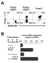
Figure 5. Rapid induction of Foxp3 mRNA by IL-2
(A) Induction of Foxp3 is not associated with cell proliferation. Purified CD25lo or CD25hi Foxp3, and Foxp3+ CD4SP cells were labeled with DDAO-SE and cultured in the presence of 5 U/ml human IL-2 and 5 ng/ml IL-7 for 24 hours. Dilution of DDAO-SE and acquisition of Foxp3 was assessed by flow cytometry. DDAO-SE staining after proliferation is shown in Suppl. Figure 8. (B) Analysis of Foxp3 mRNA induction by real time RT-PCR. Purified CD25hi Foxp3 CD4SP cells were cultured with or without 5 U/ml human IL-2 for either 3 hours, or for the last 3 hours of a 6 hour culture, and assessed for Foxp3 mRNA expression using real time RT-PCR normalized to HPRT expression (Hori et al., 2003). For reference, freshly isolated Foxp3+ thymocytes express 3.6 normalized amount of Foxp3 mRNA.
Figure 6
Figure 6. IL-2 and IL-15 are the predominant common γ-chain cytokines which induce Foxp3
(A) Analysis of common γ-chain cytokines for their ability to induce Foxp3. Sorted CD25hi Foxp3 CD4SP cells were assayed with the indicated murine cytokines (100 ng/ml) and IL-7 (5 ng/ml) as described in Figure 4A. Titration of cytokines down to 1 ng/ml did not enhance the percentage of Foxp3+ cells observed (data not shown). (B) IL-2 is more potent than IL-15 for the induction of Foxp3. Purified CD25hi Foxp3 CD4SP thymocytes were cultured with IL-7 (5 ng/ml) and indicated concentrations of murine IL-2 or IL-15 and analyzed for Foxp3 expression by flow cytometry at 24 hours. Data is representative of two independent experiments. The difference in potency between IL-2 and IL-15 was calculated using the concentration at half maximal Foxp3 induction.
Figure 7
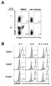
Figure 7. Upregulation of proximal IL-2 signaling correlates with the ability to induce Foxp3
(A) Foxp3 induction by IL-2 is Jak kinase dependent. Purified CD25hi Foxp3 CD4SP cells were cultured with or without 5 U/ml human IL-2 in the presence of Jak inhibitor I or the carrier (DMSO) only. Foxp3 expression was assessed by flow cytometry at 24 hours. (B) CD25hi, but not CD25lo, Foxp3 CD4SP thymocytes are capable of generating proximal IL-2 signals. Purified cells were incubated with serum free DMEM for 10 minutes, stimulated with 1000 U/ml human IL-2 or 100 ng/ml IL-7 at a concentration of 5x10⁵ cells/ml for 20 minutes, then fixed and stained for phospho-Stat5 (pStat5) as previously described (Van De Wiele et al., 2004), and analyzed by flow cytometry. FACS plots are representative from three independent experiments. Shaded area and solid line represent unstimulated and stimulated cells, respectively.
Figure 8
Figure 8. Ability to signal via IL-2 does not define the cytokine responsive Treg cell precursor
(A) Biphasic expression of CD122 on CD25hi Foxp3 CD4SP thymocytes. Data shown are gated on CD4SP cells. The bottom histogram represents the sort gating for the CD25hi Foxp3 cells of the top and bottom quartile based on CD122 expression. (B) Almost all CD122hi CD25hi Foxp3 cells have the capacity to signal via the IL-2 receptor. Analysis of phospho-Stat5 induced by IL-2 on the indicated cell subsets was performed as described in Figure 7B. (C) Induction of Foxp3 does not correlate with the ability to signal via the IL-2 receptor. Sorted cells used in (B) were also stimulated with 50 U/ml human IL-2 and 5 ng/ml IL-7 to induce Foxp3 expression, which was analyzed by flow cytometry at 24 hours. (D) Fractionation of the CD25hi subset using CD127 (IL-7Rα) and CD122 (IL-2Rβ). Flow cytometric data shown are gated on the top labels and on CD4SP cells. (E) Correlation of CD127 and CD122 with Treg cell precursor frequency in the Foxp3 CD25hi subset. CD25hi Foxp3 cells were purified on the basis of CD127 and CD122 shown in (D) and stimulated with 5 ng /ml IL-7 and 50 U/ml human IL-2 for 24 hours.

References

    1. Apostolou I, Sarukhan A, Klein L, von Boehmer H. Origin of regulatory T cells with known specificity for antigen. Nature Immunol. 2002;3:756–763. - PubMed
    1. Bassiri H, Carding SR. A requirement for IL-2/IL-2 receptor signaling in intrathymic negative selection. J Immunol. 2001;166:5945–5954. - PubMed
    1. Bayer AL, Yu A, Malek TR. Function of the IL-2R for Thymic and Peripheral CD4+CD25+ Foxp3+ T Regulatory Cells. J Immunol. 2007;178:4062–4071. - PubMed
    1. Bennett CL, Christie J, Ramsdell F, Brunkow ME, Ferguson PJ, Whitesell L, Kelly TE, Saulsbury FT, Chance PF, Ochs HD. The immune dysregulation, polyendocrinopathy, enteropathy, X-linked syndrome (IPEX) is caused by mutations of FOXP3. Nature Genet. 2001;27:20–21. - PubMed
    1. Burchill MA, Yang J, Vogtenhuber C, Blazar BR, Farrar MA. IL-2 receptor beta-dependent STAT5 activation is required for the development of Foxp3+ regulatory T cells. J Immunol. 2007;178:280–290. - PubMed

Publication types

MeSH terms

Substances