. 2008 Jan;28(1):112-21.
doi: 10.1016/j.immuni.2007.11.022.

Linked T cell receptor and cytokine signaling govern the development of the regulatory T cell repertoire

Affiliations

Linked T cell receptor and cytokine signaling govern the development of the regulatory T cell repertoire

Matthew A Burchill et al. Immunity. 2008 Jan.

Abstract

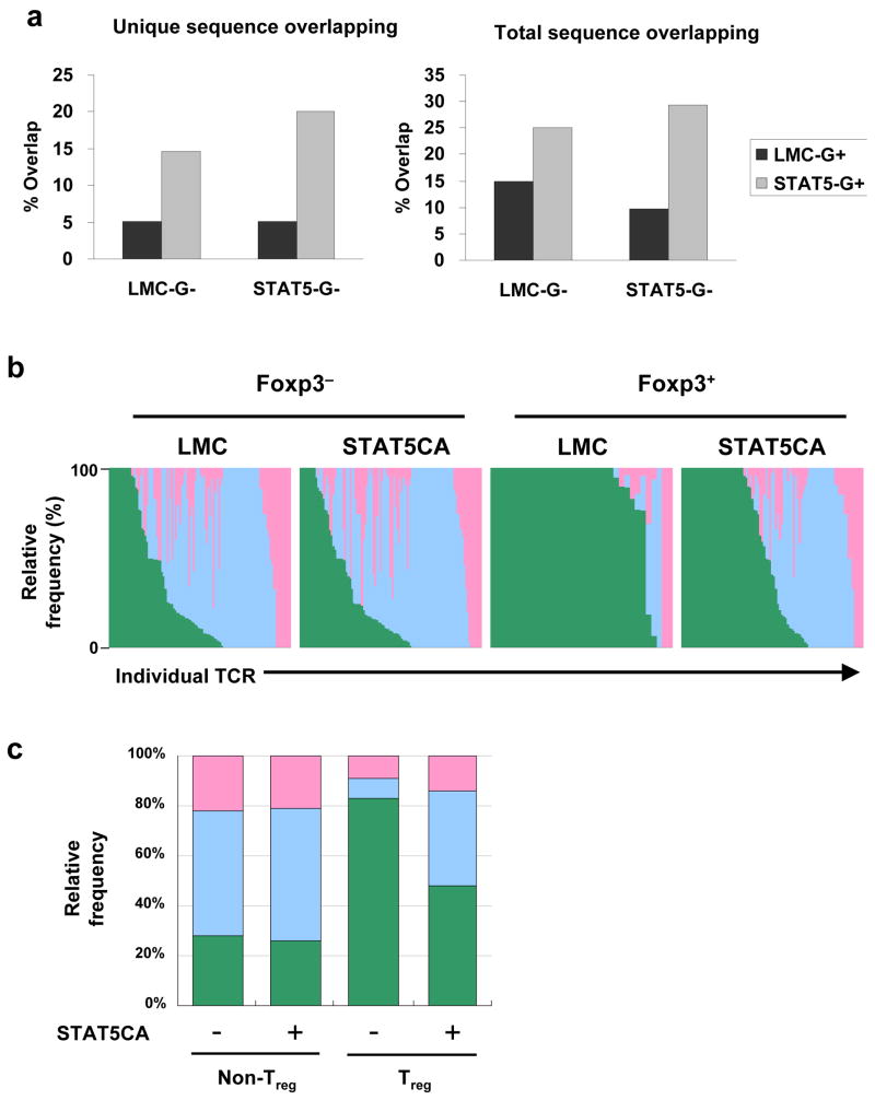
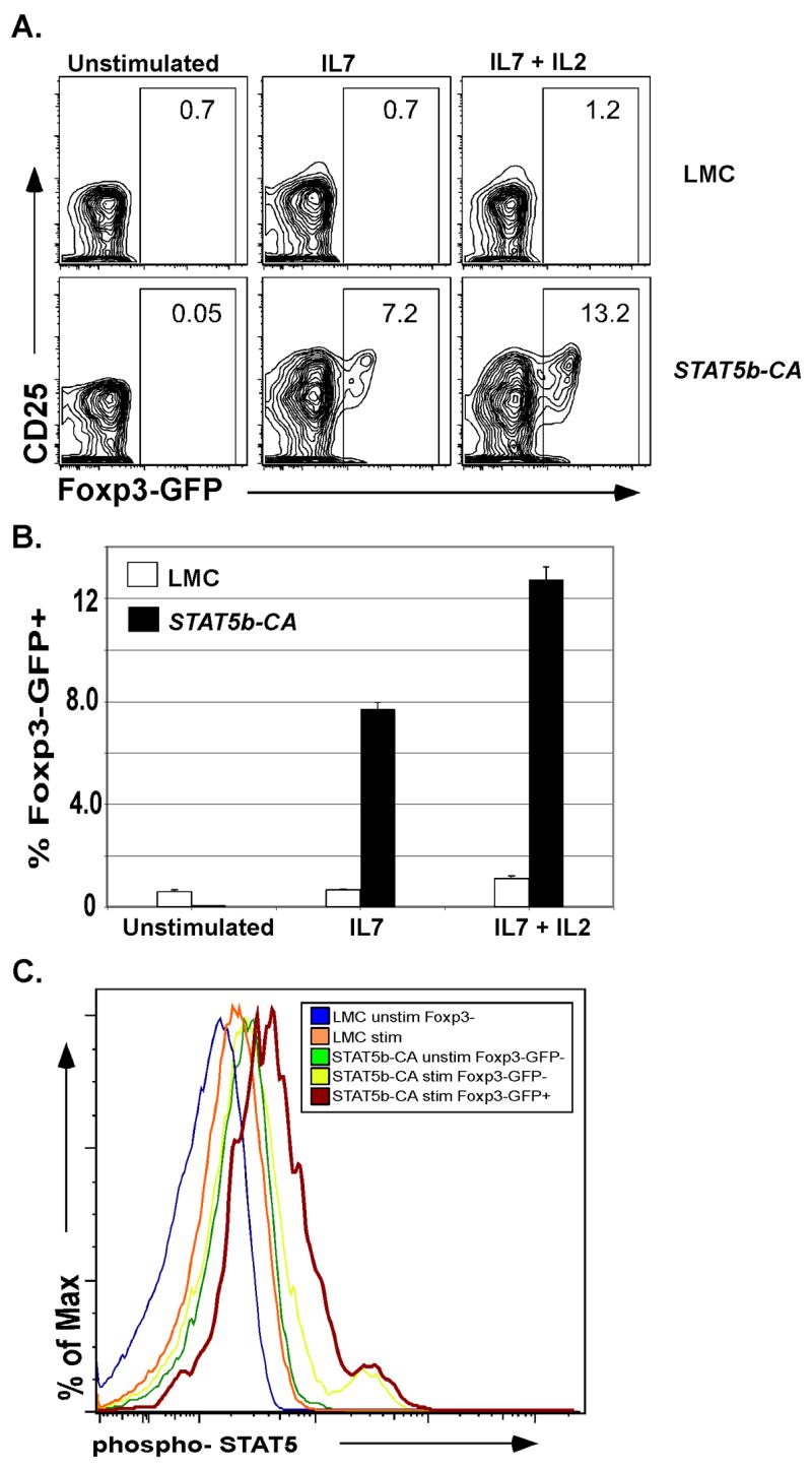
Appropriate development of regulatory T (Treg) cells is necessary to prevent autoimmunity. Neonatal mice, unlike adults, lack factors required for Treg cell development. It is unclear what these missing factors are. However, signals emanating from the T cell receptor (TCR), the costimulatory receptor CD28, and the family of gammac-dependent cytokine receptors are required for Treg cell development. Herein we demonstrate that expression of a constitutively active Stat5b transgene (Stat5b-CA) allowed for Treg cell development in neonatal mice and restored Treg cell numbers in Cd28(-/-) mice. Sequence analysis of TCR genes in Stat5b-CA Treg cells indicated that ectopic STAT5 activation resulted in a TCR repertoire that more closely resembled that of naive T cells. Using MHCII tetramers to identify antigen-specific T cells, we showed that STAT5 signals diverted thymocytes normally destined to become naive T cells into the Treg cell lineage. Our data support a two-step model of Treg cell differentiation in which TCR and CD28 signals induce cytokine responsiveness and STAT5-inducing cytokines then complete the program of Treg cell differentiation.


Figures

Fig. 1
Fig. 1
STAT5b-CA expression drives early Foxp3+ Treg development. Neonatal mice were euthanized less than 16 hours after birth. Thymii were then harvested, enumerated and stained with the cell surface antibodies CD4, CD8, CD25, HSA, and intracellular Foxp3 for analysis by flow cytometry. Similar results were found using Foxp3-GFP reporter mice. Data is representative of 3 independent experiments with 7 to 10 mice per group.
Fig. 2
Fig. 2
Constitutive activation of STAT5 drives Foxp3+ Treg development in the absence of TCR/CD28 stimulation. (A) Thymii were harvested from 4-6 week old C57Bl/6, STAT5b-CA x γc/ and γc/ mice or (B) STAT5b-CA x CD28/ and CD28/ mice and stained with antibodies to the cell surface markers CD4, CD8 and intracellular Foxp3. Numbers indicate the percentage of CD4+CD8 thymocytes (Thy) inside the gate. (C) Numbers of CD4+CD8Foxp3+ cells from LMC (white slashed bars), CD28/ (black bars) and STAT5b-CA x CD28/ (white bars) mice were calculated from total lymphocyte numbers and flow cytometry analysis in part A. Error bars represent standard error of the mean. Data is representative of three independent experiments using a total of 6–8 mice per group. (D) Shown is CD25 (left panel) or GITR (right panel) expression on CD4+Foxp3+ thymocytes from LMC (gray shaded histogram), CD28/ (blue line) or STAT5b-CA x CD28−/− mice (red line).
Fig. 3
Fig. 3
STAT5 activation promotes the development of 2W1S-specific Foxp3+ Tregs. (A) Spleens and lymph nodes were isolated and pooled from LMC or STAT5b-CA mice. Following magnetic bead enrichment cells were stained with the surface molecules CD3, CD4, CD8, CD25 and the lineage depletion antibodies CD11b, CD11c, NK1.1, GR-1, and B220. CD3+CD4+ cells were then analyzed for 2W1S and CD25 (top panels) or intracellular Foxp3 expression (bottom panels). (B) Bar graphs represent the percent Foxp3+ cells among the 2W1S (black bars) and 2W1S+ (white bars) populations. Error bars represent standard error of the mean; p-values were calculated using the two-tailed students t test. Data is representative of five independent experiments utilizing a total of 6 LMC and 7 STAT5b-CA mice.
Fig. 4
Fig. 4
Constitutive STAT5 activation alters the TCR repertoire of thymic Foxp3+ Tregs. A. Shown is the percentage of unique (left panel) or total (right panel) TCR Vα2 sequence overlap of LMC CD4+Foxp3 (LMC G-) or STAT5b-CA CD4+Foxp3 (STAT5-G-) with CD4+Foxp3+ Tregs from either LMC (black bars) or STAT5b-CA (gray bars) mice. B,C. Shown is the relative frequency with which a particular TCR Vα2 sequence from either LMC or STAT5b-CA thymocytes are found in either CD4+CD44loFoxp3(blue), CD4+CD44hiFoxp3 (pink), and CD4+Foxp3+ (green) peripheral T cells. CD4+Foxp3+ thymocytes from STAT5b-CA mice exhibit an increase in the frequency with which TCR Vα2 sequences are also found in the naïve CD4+CD44loFoxp3 repertoire (see expanded blue area (B) or bars (C)).
Fig. 5
Fig. 5
Constitutive STAT5 activation drives CD4+Foxp3CD25 thymocytes into the Foxp3+ Treg lineage in vivo. A. Shown is a representative example of Foxp3 expression among purified CD4+Foxp3CD25 thymocytes from either LMC (top) or STAT5b-CA (bottom) mice following 24 hour in vitro stimulation with either media alone, IL7 (5ng/ml) or IL7 plus IL2 (10u/ml). B. Bar graph represents the percent of cultured cells from either LMC (white bar) or STAT5b-CA (black bar) mice that induced expression of Foxp3 following in vitro culture. Data is representative of two independent experiments involving three samples per condition. C. CD4+Foxp3-GFPCD25 thymocytes from LMC and STAT5b-CA mice were placed in culture overnight with or without IL2 and IL7. Cells were subsequently stained for phospho-STAT5 expression and analyzed by flow cytometry. Shown are relative phospho-STAT5 expression for unstimulated CD4+Foxp3-GFP LMC cells (blue), unstimulated STAT5b-CA CD4+Foxp3-GFP cells (green), stimulated LMC CD4+Foxp3-GFP cells (orange) and stimulated STAT5b-CA CD4+Foxp3-GFP cells (yellow) and CD4+Foxp3-GFP+ cells (red). A representative example of two experiments is shown.
Fig. 6
Fig. 6
A. TGFβ signaling is not essential for conversion of CD4+Foxp3-GFPCD25 thymocytes from STAT5b-CA mice into Foxp3+ Tregs. CD4+Foxp3-GFPCD25 cells from STAT5b-CA mice were stimulated for 24 hour in vitro with either media alone (left panel), IL2 (10 U/ml), IL7 (5ng/ml) and 10 μg/mL isotype control antibody (middle panel), or 10μg/mL anti-TGFβ antibody (right panel). B. Histone deacteylase activity is required for cytokine/STAT5 dependent conversion of CD4+ thymocytes into Foxp3+ Tregs. CD4+Foxp3-GFPCD25 thymocytes from STAT5b-CA mice were stimulated overnight with IL2 (10 U/mL) plus IL7 (5 ng/mL) and either 0.2% DMSO (left panel), 100 nM trichostatin A (TSA, middle panel), or 800 nM apicidin (right panel). Twenty-four hours later cells were stained for CD4 expression and analyzed for Foxp3-GFP expression by flow cytometry. Shown is a representative example of three experiments.

Similar articles

Cited by

References

    1. Anderson MS, Venanzi ES, Klein L, Chen Z, Berzins SP, Turley SJ, von Boehmer H, Bronson R, Dierich A, Benoist C, Mathis D. Projection of an immunological self shadow within the thymus by the aire protein. Science. 2002;298:1395–1401. - PubMed
    1. Apostolou I, Sarukhan A, Klein L, von Boehmer H. Origin of regulatory T cells with known specificity for antigen. Nature immunology. 2002;3:756–763. - PubMed
    1. Aschenbrenner K, D’Cruz LM, Vollmann EH, Hinterberger M, Emmerich J, Swee LK, Rolink A, Klein L. Selection of Foxp3+ regulatory T cells specific for self antigen expressed and presented by Aire+ medullary thymic epithelial cells. Nature immunology. 2007;8:351–358. - PubMed
    1. Baldwin TA, Hogquist KA. Transcriptional analysis of clonal deletion in vivo. J Immunol. 2007;179:837–844. - PubMed
    1. Bassiri H, Carding SR. A requirement for IL-2/IL-2 receptor signaling in intrathymic negative selection. J Immunol. 2001;166:5945–5954. - PubMed

Publication types

MeSH terms